TOTAL HIP REVISION AT SIRIRAJ HOSPITAL
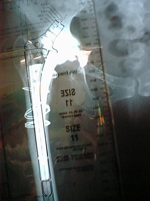
Fig 02: Pre-operative x-ray with size template iver it to asses the size of the prosthesis
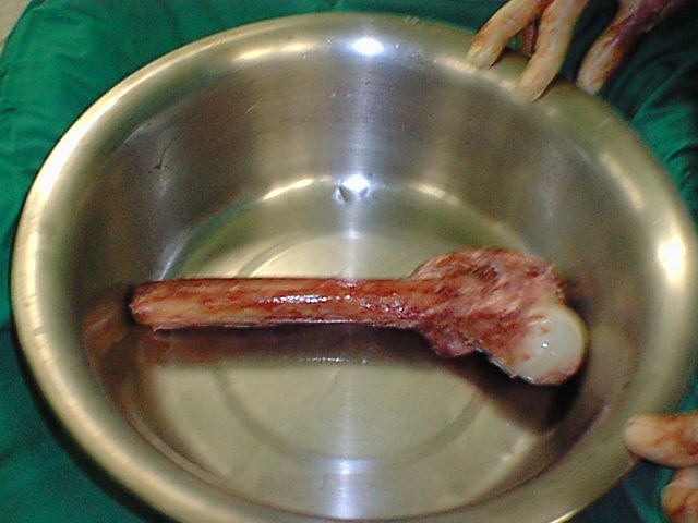
Fig 03: Prosthesis removed from femur
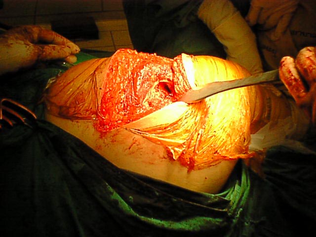
Fig 04: Femoral canal being reamed and cleaned
Fig 05: X-ray of the femur allograft selected for substituting the defect
Fig 06: View of femur allograft
Fig 07: Measurement of Calcar defect with the help of femoral trial
Fig 08: Size of allograft to fil the Calcar defect being taken
Fig 09: Reaming of the canal for Calcar sizing and prosthesis fitting
Fig 10: Sizing of the Calcar allograft with the orignal prosthesis
Fig 11: Freeze dried Cancellous chips mixed with patients blood
Fig 12: Allograft and prosthesis being seated on the femur
Fig 13: Allograft being tied with circlage wire
Fig 14: Immediate post-operative X-ray of the patient
THANK YOU FOR BEING WITH US