MISCELLANEOUS
OK..... sit back, relax and have a coke.
I stumbled across some arachnid which left me clueless. Many speculations were
made ranging from the elusive ricinulei to spiders to whatever.....
Thanks to response from Dr Peter Schwendinger, Dr James Cokendolpher and Theo Blick,
the specimen concerned should be Oncopus acanthocelis ROEWER of Family
Oncopodidae from the Order Opiliones. Dr Peter Schwendinger did a recent revision of
the family in 1998. O.acanthocelis is found in South East Asia and has
fused dorsal tergites which is seen in the specimen.
The photos using a video conferencing PC camera is of bad quality and the need for
compression (used to be at least 10x the size) makes it worse.
Some background on this arachnid
Number of specimens: 2
Length from head to end of body: 6-7mm, no hoods present.
Location it was found:
A tropical rainforest, under bark of a dead fallen tree. Very moist environment.
Proximal to a centipede probably a mid sized Scolopendra subspinipes and a small
termite nest (species was not determined then)
Number of legs: 4 pairs
Second pair of legs elongated. 1st pair of legs shortened.
one pair of palp present (just lateral to the chelicera like appendages)
One pair of large chelicerae present.
Behaviour: Most active at night. Shy to light.
Motion slow and ponderous. AVI not available online but a compressed version of 900kb can
be downloaded from me.
Does not show any aggressive tendency to each other or external stimulus.
Diet: Thanks to help from R. G. Breene, I attempted feeding the specimens 2
different species of termites. Saw one being consumed by specimen 1 but
unfortunately, no recording was done. Rest of the termites (all 8 of them)
disappeared overnight. Even when moving to prey, no change in pace was observed. The
specimens seems immune to biting and acid attack of soldier termites. The angle of
the chelicera motion and the 'hugging' of the prey with the palps resembles that of
Theraphosids. No further predation was seen 'live'.
Below are the pictures (14 pictures)
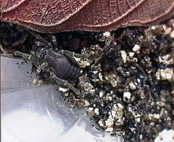
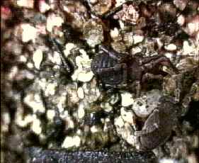
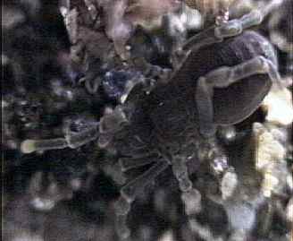
Specimen 1: Looks like a freshly moulted individual.
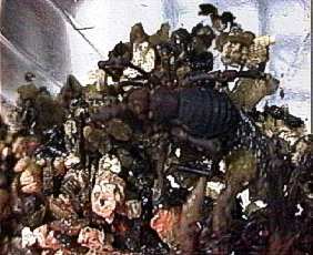
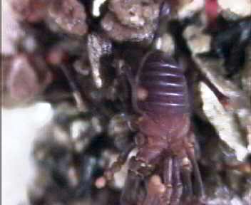
Specimen 2. The posterior region seems segmented.
The constriction between the 'head' and the 'abdomen'
does not seem to be a movable joint.
A 45 degree shot of specimen 2
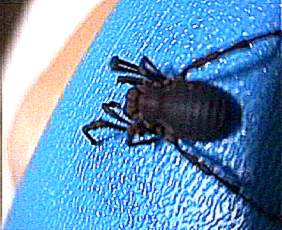
Specimen 2 from side
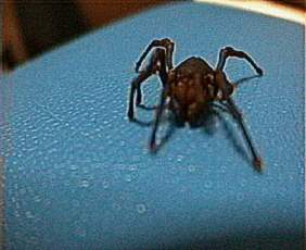
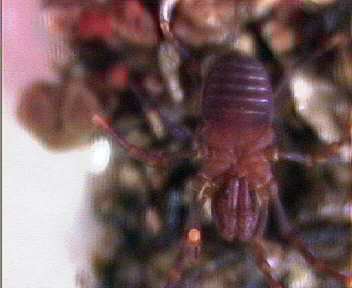
Frontal view of specimen 2
Anal view of specimen 2. No spinnerets is seen
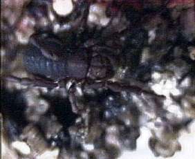
Specimen 2 illustrating the well marked chelicerae
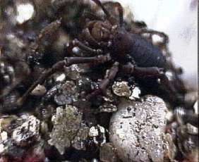
Both specimens seen in this shot. Note that one
of them is of much lighter colouration. Specimen 2
on top. Specimen 1 below.

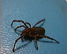
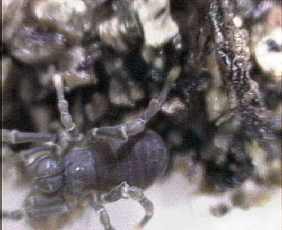
A close up of the specimen 1. Note the huge
pair chelicerae.
Another closeup of specimen 1.
A clearer shot of specimen 1 showing some marking
on the chelicerae.
Specimen 2.
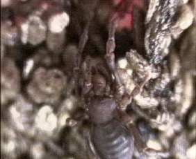
Ventral view of the specimen 2. Note that the palps extends
posteriorly to the coxa 2. Its tip is black in color. Segmentation
of the body is clearly illustrated.
Straight on ventral shot of specimen 2. The tarsus seems to
be rounded. Original colour of specimen.