Three Cases of a Hitherto Unclassified Affection
Resembling in its Grosser Aspects
Obesity, but Associated with Special Nervous Symptoms --
ADIPOSIS DOLOROSA
By F. X. Dercum M.D.
CLINICAL PROFESSOR OF DISEASES OF THE NERVOUS SYTEM IN THE JEFFERSON
MEDICAL COLLEGE; NEUROLOGIST TO THE PHILADELPHIA HOSPITAL.
At the meeting of the American Neurological Association held in Washington in September, 1888, the writer reported an anomalous case found in the nervous wards of the Philadelphia Hospital, and as it was not possible to classify the condition found, the description was prefaced by the title, " A Subcutaneous Connective-tissue Dystrophy of the Arms and Back associated with Symptoms resembling Myxœdema." Subsequently the case was published in the University Medical Magazine for December, 1888.
Some two years later, another and apparently similar case was discovered in the medical wards of the Philadelphia Hospital, and was reported at a meeting of the Philadelphia Neurological Society in December, 1890, by Dr. Frederick P. Henry. It was described as a case of myxœdematoid dystrophy and afterward-published in the Journal of Nervous and Mental Disease for March, 1891. Dr. Henry stated that he adopted the term dystrophy in order to bring the case "into the same category with the very similar one" reported by the writer.
In October, 1891, a third case, resembling the others, made its appearance in the nervous wards of Blockley. This will presently be detailed, but before doing so let us review, as briefly as possible, the previous cases, in order that all three may be considered together.
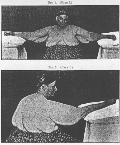
Case I. -- Before described by the writer, as stated, in 1888.
History in abstract as follows: -
M. G., aged fifty-one, female, widow, native of Ireland, domestic.
Family history. Father died at forty-five, of erysipelas. Mother had eighteen children; died at forty from some complication incident to the menopause. Of brothers and sisters, seven died in early childhood, one in adult life, of pleurisy; a sister, in childbirth; a brother and two sisters, of phthisis; while the remaining five are living and apparently in average health. None of the patient’s relatives had ever suffered, as far as she knew, from symptoms similar to her own. No history of insanity, epilepsy, or other neurosis.
Personal History: As a child, had measles, whooping cough, and scarlet fever. Menstruated normally at fifteen. Married at eighteen. Some years after, had an attack of pneumonia but made a good recovery. Had in all seven children and one miscarriage. Five children died in in-fancy or childhood, one from cholera infantum, two from measles, one from " congestion of the brain," and the fifth from " spasm." Menopause set in abruptly at thirty-five. From this time up to within two or three years her health had continued good. She had undergone some increase in weight, but beyond this nothing worthy of mention could be recalled. Syphilis and alcoholism denied.
When forty-eight or forty-nine years of age noticed that her arms were becoming very large. The upper arms and shoulders appeared swollen. On some days the swelling seemed more decided than on others. it continued steadily to increase, and for about a year was unattended by any other symptom.
In November, 1886, she was admitted to the surgical wards of the Philadelphia Hospital for the rupture of a -varicose vein of the leg. In the following February she was transferred to the medical wards for a severe -attack of bronchitis. Later she had an attack of severe pain and swelling in the right knee, attended by chill and fever. She was treated for rheumatism and promptly relieved. Two weeks after this she complained of a sharp darting pain in the right arm. It began on the outer aspect above the elbow, and gradually increased in severity and extent, spreading upward to the shoulder and neck and downward to the forearm and hand. It was different, she states, from the pain previously experienced in the knee. It was shooting and burning. She felt at times as though hot water were being poured upon the arm, and again as though the hand and fingers were being torn apart. No rise in temperature was noted. The pain was often paroxysmal, being very much worse for hours and days at a time, but even during the intervals it was never altogether absent. On June 4, 1887, she was removed to the nervous wards, when she came under the writer’s care.
Her appearance at this-time was striking. She was a tall, large-framed woman who looked as though she had at one time presented a fine physical development, but she seemed unnaturally broad across the back and shoulders. On removing the clothing an enormous enlargement of-these parts was disclosed. The enlargement affected both shoulders, the arms, the back, and the sides of the chest. It was most marked in the upper arms and back, forming there huge and somewhat pendulous masses, It was elastic, and yet comparatively firm to the touch, and it was impossible to produce pitting. In some situations it felt as though finely lobulated, and in others, especially on the insides of the arms, as though the flesh were filled with bundles of worms. The sensation to the fingers was very much like that experienced in examining a varicocele, except that the structures appeared more resistant. The skin itself was evidently not thickened. It did not take any part in the swelling and it was not adherent to the subjacent tissues. It was slightly roughened over the forearms, less so on the arms, shoulders, and hands, while over the fingers it was quite smooth and even glistening. Further, over the forearms and hands it was slightly darkened, small brownish patches and minute epithelial scales being observed; lastly it was quite dry.
The right arm was extremely painful on motion. The head, at this time, was also held in a fixed position for fear that movement of the neck would give rise to pain in the shoulder. In addition, the arm was also very sensitive to pressure. Pronounced pressure appeared to be absolutely unbearable. The nerve trunks also were exquisitely sensitive, but this painful condition was not by any means limited to them, but permeated the swollen tissues as a whole.
In marked contrast to the right, the left arm could be handled with impunity. Transient pain was, however, at one time noted in the left wrist.
The muscles were evidently not involved in the swelling. On grasping, for instance, the enlargement over the left biceps and directing the patient to flex and extend the arm repeatedly, the mass was felt to be unaffected by the movements of the underlying muscle. The affected parts were, however, quite, weak. The grip of the right hand was almost nil, while that of the left was greatly diminished. Examined electrically, the muscles of the shoulders and arms yielded a negative result, partly because of the great resistance caused by the intervening tissue. Slight quantitative and qualitative changes were noted in the muscles of the forearms, while in the hands distinct reaction of degeneration was noted in the thenar and hypothenar groups, more evident on the right side.
Cutaneous sensibility was much diminished. On the right arm various areas existed in which no response whatever was given to the aethesiometer. They were large and irregular in shape and very sharply ,defined, and were present on both the inner and outer aspects In the finger-tips of the same side the points could not be at all separated. In the left arm, on the other hand, the response was prompt and accurate, except on the outer aspect of the forearm, where some delay and uncertainty existed. In the finger-tips, also, sensation was decidedly below normal, the points being separated at not less than one-half or three-quarters of an inch, Sensibility to heat and cold was also diminished.
Examining the legs, it was found that cutaneous sensibility was distinctly lessened on the right, while showing little or no impairment on the left. No enlargement was, however, noted at this time in any portion of the body, save in the regions already mentioned. No swelling or anćsthesia was found about the face. The latter was pale, as were also the mucous membranes. There was, however, a little color in the cheeks, more noticeable at times. Her features were well formed and intelligent. Her hair was dark and fine, and somewhat thin over the vertex. Her mind was unimpaired, except that at times she was much abstracted. Sometimes she gave conflicting answers to questions, so that the latter had often to be repeated. Her speech was not slowed or otherwise altered. At times she was irritable and quarrelsome, and frequently gave much trouble to her nurses.
The above abstract fairly represents her condition at the time of her admission to the nervous wards.
June 13th, ten days later, she had a chill, followed by fever and a painful herpetic eruption over the upper portion of the left arm and the upper and anterior portion of the left side of the chest. June 19th, another crop of vesicles made its appearance on the back and on the front of the chest.
For some three months following, among other studies, a careful record of the axillary temperature was made. It proved to be very nearly normal. At one time, however, a temperature of only 97° was recorded.
Nothing worthy of note occurred until October 13th, when the patient had another severe attack of bronchitis, which was accompanied by much dyspnœa.
In the latter part of December it was noticed that during one of her paroxysms of pain the swelling of the right arm became more decidedly lobulated. The arm became more sensitive than ever, and on examination hard, cake-like masses were felt, resembling, as the resident physician expressed it, the caking of milk in a breast. This caking, or increased lobulated feel, was subsequently repeatedly noticed during paroxysms of pain. At this time, also, she suffered from an attack of pain in the right knee, and in the popliteal space a diffused swelling was felt which exhibited the same curious nodulated or leech-like feel as did the swelling elsewhere. It was also very painful, but subsided in a few days, and no permanent alteration of the tissues could be detected.
At various times subsequently paroxysms recurred, during one of which swelling was noticed in the posterior triangles of the neck, which seemed later to be permanently fuller than normal. Bronchitis also recurred, accompanied by dyspnœa, and at one time with free expectoration of bloody mucous.
In April following she experienced an attack of unusual severity. The pain, which involved the right arm and shoulder, right side of trunk and back of neck, now for the first time spread to the face and head. The right side of the face and neck became distinctly swollen, and presented to the touch the same nodulated feel so characteristic of the swelling in other portions. At the same time, the tongue and probably the pharyngeal tissues became swollen. Her tongue, she said, felt much too large for her mouth, and this certainly appeared to be the case. Her speech was much interfered with. Her voice was very hoarse, and she spoke with great difficulty. This condition persisted for upward of a week, when the swelling slowly subsided. For some time subsequently she spat blood, the source of which was not determined, though it appeared to come from the throat. The reddish color in the cheeks also became more pronounced, until it covered the entire forehead like an intense blush. This blush was afterward observed to recur with other paroxysms of pain.
During the summer of 1888 the patient’s condition underwent some change. The paroxysms of pain became less frequent and less severe. Hand-in-hand with this improvement, sweating became very abundant. However, paroxysms accompanied with marked dryness of skin occurred from time to time, and upon one occasion a thick, welt-like swelling, exquisitely painful, was observed extending from the upper and inner angle of the scapula perpendicularly down the back to very nearly the lumbar region. Upon another time, swelling again made its appearance in the right popliteal space, as well as on the inner aspect of the knee. In the latter locality the swelling became permanent and the tissue presented the same peculiarities as noted elsewhere.
Pain now occasionally appeared in the left arm. Prolonged attacks of cardiac dyspnœa recurred every week or two, and apparently independently of bronchitis.
Examination of the eyes by Dr. de Schweinitz revealed contraction of the fields of vision for form and colors, most marked in left eye. The other special senses, hearing, taste, and smell appeared to be somewhat obtunded.
An analysis of the urine yielded a negative result. A blood-count failed to reveal an increase of white corpuscles.
Since the notes from which the above account is condensed were taken, the patient has at various times during attacks of pain vomited blood. Upon several occasions this was observed by the writer himself. The quantity could not be accurately estimated, but while it was never in large bulk at a single emesis it was constantly brought up in repeated vomiting during an entire night or day. The last attack occurred in August, 1891.
Measurements were made of this patient at various times, and there has been a steady increase in the bulk of the arms up to the present time.
As a whole, however, the patient has not suffered as intense pain as formerly. Cardiac dyspnœa, though, is a frequent and very distressing symptom. Pulse soft and rather rapid, ranging from 95 to 110. Face still flushed. Of late has had shooting pains in the abdomen, and examination discloses an extensive deposit of tissue in this region, and to which the pain is referred. A large longitudinal wheal, especially sensitive, is found in the left lumbar region.
A deposit of tissue (or swelling) has also made its appearance over the left hip and to some extent over the right. Thighs and buttocks do not seem to be especially enlarged, but soft masses are now found on the .inner sides of both knees, the right larger than the left, the former more painful to pressure.
A small nodule to the right of the scorbiculus is especially painful.
At various times, by means of a Duchenne trocar, fragments for microscopic examination were removed from either arm. They revealed connective tissue and fat cells present in varying degree. It was observed that the former was decidedly embryonal in type, the cells being large and fusiform, and their nuclei correspondingly large and prominent. The fat-cells for the most part were associated with these connective-tissue cells, and occasionally individual fat-cells were seen in which the fatty metamorphosis had not been complete. (In one of the fragments removed the writer was fortunate enough to find nerve elements which had probably been included in the grasp of the trocar by the latter grazing a blood vessel, as the fibres were non-medullated. Their connective tissue was denser than normal and they presented an unusual number of nuclei.) For a detailed description, with drawings, the reader is referred to the original article (loc. cit.).
CASE: II. -- This, as already stated, was reported Dr. F. P. Henry (loc.
cit.). Subsequently in November, 1891, she came under the care of the writer, having
been transferred to the nervous wards, where she finally died about a month later. The
following account is abstracted partly from the report of Dr. Henry and partly from my own
notes.
E.W., aged sixty-four, married, native of England.
Family history. Father dead of alcoholism at middle life. Mother dead at twenty-eight of œdema of brain, verified post-mortem. Has living an elder brother and sister and one younger brother. The younger brother when a child was "peculiar" -- he would run to people in sudden fright and say that he was drowning or the like. He is now in average health, but drinks heavily. Has a contracture of the ring finger. Has nine children, all of whom appear to be well. The older brother suffers periodically from violent headache; also, since a young man has suffered from constantly cold feet -- this so severe as to disturb sleep and cause great distress. He had five sons and two daughters. One son died of tetanus (traumatic) ; the others are well. One daughter has a contracted middle finger of the right hand; has never suffered pain in the finger. Patient’s sister is living, sixty-five years old, and healthy; no children.
Previous history. Does not remember having the ordinary diseases of childhood. At early infancy began to have fits, which at times occurred daily, at other times weekly. Consciousness was lost during the fits, and they were followed by great pain in the forehead. After the seizures she slept. During this time was relieved of lumbricoid worms -- vomiting them; and some time later recovered from the fits.
Was married at seventeen. Had two sons, the older of whom is now forty and has seven healthy children; the younger died at two years of hemorrhagic diarrhea. Patient bad no miscarriages and no stillbirths. Left her husband because of a venereal disease contracted by the latter. Was told by a doctor that she had escaped infection. A year later, however, she had sore-throat with white patches. Had been an immoderate drinker for many years. For weeks at a time was intoxicated every night.
Menstruation began at eleven and ceased abruptly at thirty-five. Lost habitually an unusual quantity of blood, but never suffered any discomfort.
History of present disease. Her malady began about fifteen years ago, when she was forty-nine years old. At that time she was living in California. The first thing noticed was a constant feeling of coldness about the knees, followed by swelling, which gradually increased. At first she thought the swelling was due to her growing fat, but later was astonished to see that there was a localized mass on the Inner aspect of each knee. At the same time there was dull aching pain in the affected parts. Later the right arm became involved, a mass making its appearance on the outer aspect. Her body she now noticed also became larger, as her stays were too small for her. During this time, while still in California, her inability to perspire, except at the Turkish bath, was marked and was part of her reason for coming East. Since she has been in Philadelphia the lack of perspiration has not been as marked as before. Various plans of treatment were tried, but did not influence the progress of the disease (that is the growth of the swelling). Five or six years ago injections of chloroform were made into the swellings on the inner side of the knees, but no good was done. Painful ulcerations were the result, and scars of considerable size mark their location,
About five years since a slight swelling appeared in the epigastrium. This gradually increased in size until it resembled the breasts in shape. and afterward spread so as to involve nearly the whole abdomen.
From the knees the process extended to the thighs, and gave rise to the large masses on their outer side and about the hips.
To Dr. Henry she stated that pain had never been a well-marked feature of the disease, which differed, however, from her statements made to the writer. To both, however, she stated that at various times she had suffered with pains apparently situated in the enlarged tissues or running down the limbs. Sometimes these attacks were fairly well localized, in one limb, in one side, or about a joint.
Five years ago her attention was called to a peculiar condition of the right hand. The last phalanx of the second finger began to be fixed in a flexed position, while the end of the finger appeared to be growing somewhat smaller. Later the remaining fingers of this hand became involved and all the phalanges deformed. The deformity as seen now is flexion of first phalanx, marked over-extension of second, and half-flexion of the third. The thumb is also stiff, but all of its joints are flexed. For some time she has noticed the thumb of the left hand becoming like that of the right.
A year ago the patient had a quasi-rheumatic attack affecting the deformed hand and the arm. The pains seemed to run up and down in the arm rather than about the joints.
Some months ago had pneumonia of the right lung, and made a good recovery.
For several months past had slight uterine hemorrhage at times, associated with which were dull, aching pains, resembling those formerly felt before menstruation.
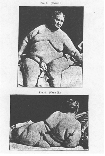
On November 27, 1891, she was transferred, as already stated, to the nervous wards. Here, on questioning her in detail, I was able to confirm the points of the history, as described by Dr. Henry. In addition, she said that the enlargement had spread from the knees to the thighs and buttocks unequally, that the left thigh and buttock had been earlier and more conspicuously enlarged than the corresponding parts on the right side. Gradually, however, the latter became enlarged to all almost equal degree. Later, swelling appeared over the left arm, and later still on the back and sides of the trunk, and wherever appearing gradually became diffused and finally reached very great proportions. The patient further volunteered the statement that she had formerly been very slight in build.
To ordinary observation she merely presented the appearance of an excessively obese person. However, examination soon revealed that the enlarged tissue was very unevenly distributed. In the region of the knees, where it had first made its appearance, it was excessively irregular and lumpy. To the fingers it resembled, in a remarkable degree, the swollen tissues of Case I. It gave the same nodular and elastic feel, and could not be made to pit on pressure. At the time of the examination no tenderness existed in any of the lumps, but shooting pains were referred to them in various situations. This was particularly the case in the mass over the right hypochondrium. In addition, she complained of scalding sensations on the inside of the right cheek and the right side of tongue. Nothing abnormal could be discovered in the mucous membrane of these parts. No tenderness existed in any of the nerve trunks at the time of the examination. The patient was excessively weak, and could move about her bed or sit up only with great difficult Her grip was almost nil. No tendon jerks could be elicited -- probably due to purely mechanical difficulties. For the same reason, an electrical examination could not be made.
Examination of the cutaneous sensibility confirmed, in general, the findings of Dr. Henry, except that some areas bad become entirely anćsthetic. Dr. Henry found that there was slight analgesia, and diminished temperature and tactile sense, and further that the "changes of sensory acuteness were not more marked over the distribution of any of the cutaneous nerves, but seemed dependent entirely upon the amount of the subcutaneous tissue." Dr. Henry, it appears, found no area of absolute anesthesia anywhere. However a year later, such an area undoubtedly existed on the back of the left arm, and extended thence over the posterior aspect of the left shoulder. On the opposite side, anćsthesia, was not present, although no marked difference, if any, existed in the amount of the subcutaneous tissue. A marked increase in this subcutaneous tissue bad, however, everywhere taken place during the past year. Comparing, for instance, the measurements of tile arms made by Dr. Henry and myself, it was found that the left forearm had increased one and seven-eighths inches, and the right forearm one and three-eighth inches; the left arm one and a half, and the right arm two inches. This Increase seemed to be maintained throughout.
Subjectively, the patient complained such of headache. Her face was very much flushed, and she suffered greatly from cardiac dyspnœa. It was a persistent and distressing symptom.
Examination of the eyes proved negative, as did also that of the urine. Perspiration, according to the patient’s statement, was scant. Face not involved in the enlargement. No subnormal temperature. Hair thin, but not excessively so. No difficulty in speech. No mental impairment.
The patient remained very much in the same condition for some two weeks following her admission to the nervous wards, when her dyspnœa, already soft and compressible, became his condition, although relieved from time and feet became puffy, the face cyanotic, and the lungs œdematous and congested. Death occurred on December 22, 1891.
Autopsy, December 23rd. Body of a very large woman. Weight estimated at about three hundred pounds. Face dark from venous congestion. Some discoloration on under surface of body and thighs. A number of large white scars on either side over the knees. Legs and feet œdematous. Body distorted and flattened, as though by its own weight.
Scalp and calvarium revealed nothing abnormal. Veins of dura and longitudinal sinus full. Venous congestion of the pia. Cortex a little darker than normal. Puncta vasculosa prominent. Brain otherwise normal. Spinal cord appeared normal. Skin of thorax appears normal. The subcutaneous tissue is fatty and moist.
Thyroid gland small, indurated and infiltrated by calcareous matter in both lobes.
Right lung œdematous and tightly adherent to chest walls. Left lung œdematous, with hypostatic congestion posteriorly. Both pleural cavities contain a large excess of fluid.
Pericardium contained some six to eight ounces of fluid, in which was suspended some flocculent lymph. Weigh of heart twenty-seven ounces; the right side dilated, the moderator band much thickened. Walls of left side also much thickened; marked hypertrophy of the columnć carneć and papillary muscles. Some fatty change, especially in walls of right ventricle.
Over the abdomen the subcutaneous fatty tissue was three inches thick. About a pint of ascitic fluid in abdomen. Stomach much dilated. Intestines normal. Liver showed some fatty infiltration, otherwise normal. Spleen apparently normal, though somewhat dark. Kidneys both reveal, except slight adhesion of the capsules, nothing specially abnormal.
In the pelvis, an ovarian cyst containing some six ounces and a hydrosalpinx were found on the left side. Uterus seemed a trifle larger than normal. Bladder normal.
Brain, cord, some of the nerve trunks, pieces of skin and subcutaneous tissue, pieces of the liver, kidneys, and spleen, a fragment of muscle, and the whole of the thyroid gland were removed for microscopic examination. The specimens were left in the care of Dr. H. W. Cattell, assistant to the pathologist of the hospital. Unfortunately, Dr. Cattell fell ill with scarlet fever, and during his absence the specimens, together with those of the next case (Case III.), were thrown away by an attendant.
CASE III. -- M. M.. aged sixty years, widowed, a tailoress by occupation, and a native of Germany, but a resident of America for twenty-six years. Admitted to the nervous wards of the Philadelphia Hospital October 7, 1891. Memory very poor. History obtained in part from relatives.
Family History. Father and mother were healthy. Mother died of heart disease. Had seven brothers and sisters, all apparently well. Had no children, no pregnancies.
Previous History. Many years ago a lump appeared at the back of the neck, for which she consulted Dr. Gross at the Jefferson Medical College, but for some reason no operation was performed. At various times thereafter swellings made their appearance in various situations.
Lost more blood at menstrual periods than normal. Occasionally suffered from hćmatemesis and epistaxis. Climacteric at forty-six. No history of any intercurrent affections. Mental impairment had been noticed for about two years.
Present condition. Patient is excessively feeble. For some two weeks vast has been unable to walk. Lies, for the most part, in a quiet apathetic state, though when aroused answers questions intelligently, but slowly. Is, in addition, somewhat deaf.
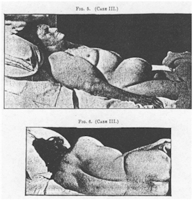
Examination reveals soft, fat-like masses or swellings in various situations. Thus a large soft mass is found over either biceps, and others, somewhat smaller, over the outer and posterior aspect of either tipper arm. Two large masses are found over the belly, separated above the umbilicus by a deep transverse crease. Another gives excessive prominence to the mons Veneris. From the back of the neck, at its lower part, springs a big mass like a hump, while a diffuse swelling gives a cushion-like coating to either half of the back, and extensive deposits give unnatural prominence to either hip. In marked contrast, the deposit is absent from the forearms and hands, from the face, front the thighs and legs and from the buttocks. The gluteal regions, in fact, seem flattened and sloping.
The deposit at the back of the neck and over the abdomen seemed tolerably firm and resistant; over other portions it was-quite soft, though elastic, and exhibited the same nodular feel noted in the previous cases. Further, it was discovered at once that these masses were painful to the touch, the patient complaining very much when only moderate pressure was exercised. This was especially true of the deposits over the arms and back of the neck. In addition, the patient complained of stabbing pains in the deposits, more marked in the regions just mentioned. There was no tenderness over the nerve-trunks. She complained also of headache.
In making the examination, it was also further noted that the left radius was rough and nodular for ;about two and a half inches in its middle third; also, that there was a large discolored area on the outer aspect of the left forearm resembling a syphilitic scar. Both tibia were somewhat nodular, though no scars were discovered on the legs. A few white scars were seen on the forehead. Quite a number of purpuric spots were also observed on the forearms, thighs, legs, and back.
The skin of the forearms and hands, and that of the legs and feet to a less extent, was dark, dry, and much roughened.
Cutaneous sensibility was found generally diminished, while a few patches of anesthesia were noted. One of these was an area diffused over the right side of the trunk and the right shoulder. They appeared to be constant, and were confirmed at various examinations.
Owing to the extreme weakness of the patient, the study of the eyes could not be made satisfactorily, but, as far as it went, was negative.
The urine contained albumin. No casts were found.
In answer to questions, the patient said that she had not been sweating freely for years, but owing to her mental condition no importance was given to this statement. She at no time presented a subnormal temperature. Her hair was well preserved.
Patient seemed to fail gradually, although diet and stimulants were freely used. Her dementia gradually deepened, and for some days before death she voided urine and feces involuntarily. She finally died in a comatose state on November 5th.
Autopsy, November 6, 1891. Body of a large woman with irregularly distributed fat-like masses. Some discoloration of the back. Small bedsores beginning on the buttocks.
Scalp and calvarium normal. Dura normal. Pia very œdematous. Brain very soft and œdematous. Cord revealed nothing abnormal.
On incising the skin of the chest and abdomen it was found to be normal in appearance, but the subcutaneous tissue, which looked like a very white fat, was excessively thick, reaching below the umbilicus a depth of seven inches.
The thyroid gland was larger than normal, harder to the feel, and much calcified, especially the right lobe.
The heart weighed eight and a half ounces. Both aortic and mitral valves were slightly thickened. Heart substance evidently fatty. The lungs were emphysematous. . The mucous membranes of the. stomach revealed a chronic gastritis. The liver weighed forty-four ounces, and beyond some fatty infiltration, was practically normal. Spleen normal. The kidneys, however, showed decided shrinking and loss of cortical substance, with somewhat adherent capsules. Nothing noteworthy in pelvic organs.
As in Case II., brain, cord, nerve-trunks skin and subcutaneous tissue, thyroid gland, and portions of other viscera were removed for microscopic examination, with the subsequent unfortunate loss of the specimens already mentioned.
It is not without some hesitation that I bring these cases before you. I am well aware that without a microscopic examination to supplement the autopsies their study is incomplete, and yet the cases are in themselves so interesting, and appear to be so unusual, that their publication in a group with such data as are at hand is more than warranted. Certainly these cases differ radically from ordinary cases of lipomatosis, and certainly the nervous symptoms present are not without a special significance. To begin, the enlarged tissue makes its appearance in a very irregular way. Nodules of soft tissue are, at first, deposited in some one situation, or perhaps in corresponding places of the upper or lower extremities. For a time the deposit is limited to these original areas, but subsequently it makes its appearance elsewhere, and may become very extensive. Regions, however, may exist which remain permanently uninvaded. In Case I. the enlargement was first noticed in both upper arms, and later in the back. Subsequently swelling made its appearance on the inner aspect of the right knee, to be followed months after by a similar swelling in a corresponding position over the left knee. Later still, it made its appearance in various other situations. However, the legs, with the exception of the knees, have remained free from involvement, while the thighs and buttocks have only recently shown a doubtful change. In Case II. the enlargement began on the inner aspect of either knee, and then gradually spread unequally over the thighs and buttocks. Later, the left arm became involved; next the sides and back, and finally the entire trunk. In Case III. the enlargement began in the back of the neck, and thence at various times in other situations. It remained absent from the face, the forearms, the legs, thighs, and buttocks. It is a peculiarity of this case, further, that the enlargement tended to produce distinct segregated masses.
Not only is the development of the enlargement irregular and even capricious in these cases, but there is, in addition, another important fact to be remembered, and that is: that at some time or other the enlargement is accompanied by pain or other nervous symptom. Thus, in Case IL, pain and a sensation of cold preceded the appearance of the nodules on the inside of the knees. In Case I. pain was noticed a year after the swelling of the upper areas had begun to show itself, and in Case III. pain was evidently present at the time of the examinations.
In Case I., again, which I had the opportunity of studying in great detail, pain was observed at numerous times. Occasionally it was observed in old areas of enlargement, and again in regions free from the latter but in which it subsequently appeared. This was especially the case in the swelling on the inner aspect of the right knee and certain welt-like formations in the back. Finally, pains, shooting or stabbing in character, were present in all cases, both at various times in the history and at the examinations. Very suggestive, indeed, were some of the paroxysms of pain observed in Case I. In some of them decided and sudden increase took place in the swelling of a part attacked, and it became, for the time being, firmer and more resistant, and occasionally more nodulated than before. Further, as already pointed out, a permanent increase or a new focus of swelling made it’s appearance. It should be remembered, too, that some of the nerve-trunks, especially those of the right arm, were very sensitive to pressure; that some of the muscles -- e.g., the thenar and hypothenar groups -- revealed reaction of degeneration, and, more significant than all, that the patient suffered on two occasions from herpes zoster.
In Cases II. and III. tenderness over the nerve-trunks could not be elicited. However, in Case I. this symptom has at present disappeared. Indeed its absence has been noted for some time past. This circumstance leads to the suspicion that Cases II. and III. were further advanced than Case I., and that the latter was really observed during a developmental period and whilst more active changes were going on.
Among the nervous symptoms must also be placed the anesthesia or diminished cutaneous sensibility already described, as well as the excessive motor weakness. It is probable that the absence or diminution of sweating also belongs, to this category. It will be remembered that this symptom was undoubtedly present in Cases I. and II., and doubtfully in Case III. We are here reminded forcibly of myxœdema, in which diminution or absence of perspiration is so prominent a symptom, and, at the same time, these cases are still further removed from ordinary obesity, in which excessive sweating is the rule. Headache was also noted in all the cases.
Among other symptoms present in these cases should be noted hćmatemesis in Case I., hćmatemesis and epistaxis in Case III., and a recurrence of uterine flow many years after the cessation of menstruation in Case III. In Cases I. and II. the menopause occurred at thirty five, and in the latter the flow was said to have been unusually free. In Case III. the menopause occurred at forty-six, and menstruation was likewise said to have been excessive. Finally, Case III. also presented a well-marked purpura. What the significance of these symptoms may be it is impossible to say. It may. however, not be out of place to recall the not infrequent occurrence of uterine hemorrhages in women who subsequently stiffer from myxœdema.
Bronchitis is a most frequent and persistent Symptom in Case I., while both Case I. and Case II. suffered markedly from cardiac dyspnœa. Both of these symptoms were absent in Case III. By their presence we are again reminded of myxœdema, in which they are frequently present.
As already stated, fragments of the enlarged tissue we’re removed from Case I. by the Duchenne trocar, as also from Case II. In both instances fat-cells and connective tissue were found in various proportions, though at times the latter was decidedly embryonal in type; this was especially so in Case I., in which in certain areas embryonal connective tissue predominated. It would seem that this is the case in the more recent formations, while in the older areas a fully formed adult fatty tissue appeared to be present. It is especially to be regretted that the loss of the specimens from the autopsies of Cases II. and III. prevented a confirmation of these results. The autopsies, however, are not without interest when it is called to mind that in both cases the thyroid gland was found indurated and much infiltrated by calcareous deposit. It is impossible, however, to correctly interpret this condition in the absence of microscopic studies.
Now, with the above data before us, what view are Ave to hold in regard to these cases? Evidently the disease is not simple obesity. If so, how are we to dispose of the nervous elements present Equally plain is it that we have not myxœdema to deal with. All of these cases lack the peculiar physiognomy, the spade-like hands, the infiltrated skin, the peculiar slowing of speech, and the host of other symptoms found in true myxœdema. It would seem, then, that we have here to deal with a connective-tissue dystrophy, a fatty metamorphosis of various stages of completeness, occurring in separate regions, or at best unevenly distributed and associated with symptoms suggestive of an irregular and fugitive irritation of nerve-trunks -- possibly a neuritis. That this, however, does not embrace the whole truth is evidenced by such symptoms as the diminished sweating, the headache, and the contraction of the visual fields noted in Case I. However, the above inference is all that we are justified in making.
Inasmuch as fatty swelling and pain are the two most prominent features of the disease, I propose for it the name Adiposa Dolorosa.
Return to the List of Articles
Return to the
Dercum's Disease Home Page
Comments about the web page format
should be sent to the Don
Please don't forget that the information provided on this site is designed to support, not replace, the relationship that exists between a patient and his or her physician.