Corticosteroid-Induced
Juxta-articular Adiposis Dolorosa
By Steven S. Greenbaum, MD, John Varga, MD
From the Departments of Dermatology (Dr Greenbaum) and Medicine (Dr
Varga),
Jefferson Medical College, Philadelphia, Pa.
Reprinted from the Archives of Dermatology
February 1991, Volume 127, pages 231-233
Copyright 1991, American Medical Association
- Long-term treatment with high doses of corticosteroids leads to the
development of truncal obesity and focal fatty deposition. These deposits
characteristically are located on the face, the nuchal and truncal areas, and
episternally, as well as in the mediastinum and epicardium. We studied a patient with
juxta-articular adiposis dolorosa who had L-tryptophan-associated eosinophilia-myalgia
syndrome and was treated with high doses of prednisone. This is the first reported case of
adiposis dolorosa occurring as a complication of corticosteroid treatment. Alterations of
fat metabolism induced by corticosteroid excess may have played a role in the development
of this unusual painful syndrome.
Adiposis dolorosa is an uncommon disorder of subcutaneous tissue that
was first described by Dercum1 in 1892 at Jefferson Medical College,
Philadelphia, Pa. The understanding of this disease has not progressed very much in the
past 98 years. It remains better understood as a clinical entity than as a physiologic or
metabolic process.
Characteristically, adiposis dolorosa is usually seen in postmenopausal
women, although younger women and, rarely, men may be affected.2 There are
multiple painful, symmetrically distributed, fatty deposits that may be either diffuse or
localized. The pain varies from discomfort on palpation to excruciating, paroxysmal
spontaneous attacks.3 Adiposis dolorosa is commonly localized to the lower
extremities (especially around the knees), where it has been termed juxta-articular
adiposis dolorosa.4 However, the painful fatty tumors of
this disease have been reported to occur in almost any location except the head and neck.5
The search for the most effective therapy for adiposis dolorosa has been
frustrating. Proposed treatments have included prostigmine and aminoacetic acid,6
non-steroidal anti-inflammatory agents,4 compression bandages,6
intravenous lidocaine,7 systemic corticosteroids, surgical excision, and
liposuction.8
We describe a patient treated with prednisone who presented with
juxta-articular adiposis dolorosa. The temporal association of high-dose corticosteroid
use and the development of the lesions, and their resolution after reduction of
corticosteroid dose, suggest a causal relationship in this case. To our knowledge, this
possible adverse effect of corticosteroids has not been previously reported.
REPORT OF A CASE
A 67-year-old white woman was referred for evaluation of painful masses
on her knees. She had been in good health until August 1989, when she noted the onset of
diarrhea, abdominal discomfort, and marked fatigue. Two weeks later she developed numbness
and swelling of her lower extremities, accompanied by weakness, tenderness, and myalgias.
She became unable to climb stairs or continue with her daily activities and was
hospitalized. She had been taking L-tryptophan (4 g/d) for 3 months for treatment of
insomnia. Her physical examination was notable for proximal muscle weakness and cutaneous
induration of the arms and legs; the hands were spared. Laboratory examination showed
striking eosinophilia (white blood cells, 3.6 x 10⁹/L). A full-thickness skin
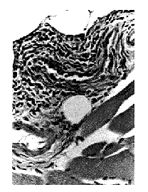
biopsy demonstrated findings consistent with eosinophilic fasciitis (Fig 1). The
L-tryptophan therapy was discontinued, and treatment with prednisone (60 mg/d) was
started. Six weeks later, because of the lack of response, the dosage of prednisone was
increased to 80 mg/d. During the following two weeks, the patient reported improvement of
myalgias, diminished swelling, and a 14-kg weight loss. Four weeks later, however, she
developed severe pain around her knees. She was referred to Thomas Jefferson University
Hospital in January 1990.
On physical examination she had full facies, oral candidiasis, a
"buffalo hump," marked truncal obesity, and grade 3/5 proximal muscle weakness.
Diffuse subcutaneous induration was present in all her extremities. Markedly tender,
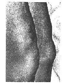
well-circumscribed, fist-sized subcutaneous masses superior to both knees were noted (Fig
2). There was no effusion in the knees. Laboratory investigation showed hyperglycemia
(glucose, 26.4) but a normal blood cell count and erythrocyte sedimentation rate. Her
serum cholesterol level was 7.75 mmol/L, and her triglyceride level was 6.42 mmol/L. A
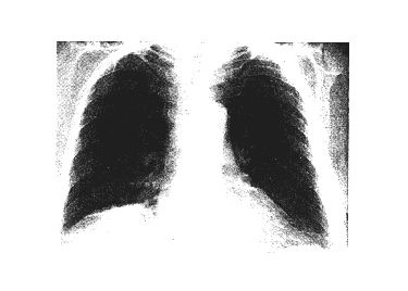
chest roentgenogram showed a widened superior mediastinum and tracheal narrowing (Fig 3).
A computed tomographic scan of the chest confirmed mediastinal widening, suggestive of
mediastinal lipomatosis. Histopathologic examination of a deep biopsy specimen from one of
the periarticular masses showed normal subcutaneous adipose tissue with a moderate
increase in vascularity. There was no evidence of inflammation, necrosis, or fibrosis
within the dermis or the septa.
During the following months, a progressive reduction of the prednisone
dose was associated with a gradual increase in muscle strength and improvement of the pain
in the knees, as well as a progressive decrease in the size of the peripatellar fatty
tumors. Four months after the patient's initial evaluation, when she was taking 30 mg of
prednisone on alternate days, these lesions had disappeared completely. A subsequent chest
roentgenogram showed a normal-sized mediastinum.

 Fig
1. --Histopathologic findings in full-thickness skin biopsy specimen of clinically
involved skin show markedly thickened fascia. There is a mixed inflammatory cell
infiltrate composed of lymphocytes, plasma cells, and eosinophils (hematoxylin-eosin,
original magnification x 100).
Fig 2.--Suprapatellar juxta-articular fatty masses. A biopsy
specimen was obtained from the mass on the right side.
Fig 3.--Chest roentgenogram demonstrating widening of the mediastinum
 .COMMENT
Our patient developed juxta-articular adiposis dolorosa while being
treated with high doses of prednisone for eosinophilia-myalgia syndrome (EMS).
Eosinophilia-myalgia syndrome is a recently described illness occurring in an epidemic
form, which develops in a proportion of individuals using L-tryptophan for the treatment
of insomnia, as well as for a wide variety of somatic complaints.9 Case control
studies from the Centers for Disease Control have firmly established an association
between L-tryptophan use and the development of EMS10 and have traced the
illness to L-tryptophan preparations produced by a single manufacturer.11
Patients with EMS have an abrupt onset of illness that is characterized by skin rash,
swelling of extremities, and severe myalgias and frequently accompanied by fever,
arthalgia, and cutaneous hypersensitivity, as well as respiratory symptoms.12
Following the stereotypic acute phase of the illness, a subset of patients with EMS
develop chronic symptoms, including muscle weakness, sclerodermalike or fasciitislike skin
changes, and neuropathy. Although cutaneous and sub-cutaneous induration is a feature of
chronic EMS, lipomas and adiposis dolorosa have not been described in these patients. No
satisfactory treatment is available for the prevention or management of the chronic
syndrome, but the use of corticosteroids in the early stages of EMS frequently results in
improvement of myalgias, tissue swelling, fever, and respiratory symptoms and resolution
of the eosinophilia.13 The role of L-tryptophan in the development of the EMS
epidemic is not well understood. This amino acid, which is available without a physician's
prescription, has been enthusiastically advocated for the management of insomnia,
depression, obesity, and premenstrual syndrome,14 and has been widely used in
the United States. No systemic illness was associated with L-tryptophan ingestion prior to
1989, but abnormalities in tryptophan metabolism have been implicated in the pathogenesis
of scleroderma15 and diffuse fasciitis.16 Adiposis dolorosa has not
been reported in association with the use of L-tryptophan.
Since the original description of adiposis dolorosa by Dercum,1
the clinical spectrum of this unusual entity has changed little. In addition to the
painful nodular depositions of fat, there are other typical components of adiposis
dolorosa. General obesity, fatigability, weakness, and a wide variety of emotional
disturbances have all been described in this syndrome. The pathogenesis of adiposis
dolorosa remains unknown. An early study suggested that a defect in the synthesis of
monounsaturated fatty acids may play a role in its development.3 Further
studies are needed to substantiate this hypothesis and to identify a specific biochemical
defect.
Long-term exposure to corticosteroids leads to gross obesity and affects
the regulation of localized fat metabolism in patients with Cushing's disease as well as
those receiving corticosteroids for disease states.17,18 Characteristically, in
corticosteroid excess, increased deposits of adipose tissue are seen along the central
body axis: truncally (usually as increased intraabdominal fit), episternally,
supraclavicularly, and in the posterior cervical area (buffalo hump). 19 The
facial fullness, termed moon facies, is due to increased buccal fat. 20
Recently, the ability of computed tomographic scanning to detect adipose tissue with great
sensitivity has led to the recognition of steroid-induced increased fat deposition in the
cavernous sinus,21 mediastinum, 20 temporal region,22 and
epidurally.23
It is sometimes difficult to determine whether fat accumulation in
corticosteroid excess is caused by in-creased localized fatty deposition or by a relative
atrophy of surrounding tissue such as muscle. It has been shown, for example, that in many
cushingoid patients there is marked thigh muscle atrophy, which tends to accentuate
truncal obesity. In addition, paraspinal and shoulder muscle atrophy makes the buffalo
hump more apparent.20
The mechanism by which steroids alter fat metabolism remains poorly
understood. Patients with Cushing's syndrome, or those who become cushingoid be-cause of
medication, display typically altered lipid metabolism, including increased levels of
very-low-, low-, and high-density lipoproteins.17,18,24,25 There are other
theories to account for the unusual distribution of fat in states of corticosteroid
excess, e.g., that it is induced by different hormones.26 Furthermore, there
appear to be regional differences in the ability of cellular receptors to bind
corticosteroids.26
To our knowledge, there are no previous reports of an association
between corticosteroid ingestion and the development of adiposis dolorosa. Our patient
developed gross truncal obesity, moon facies, and mediastinal widening due to epicardial
fat deposition, as well as markedly painful periarticular localized fatty deposits, after
taking high doses of prednisone for the treatment of EMS. These changes were suggestive of
a wide-spread dyslipoproteinemia induced by corticosteroids. She also developed diabetes
mellitus. As the dosage of prednisone was gradually lowered, dramatic improvement of
adiposis dolorosa, as well as a return to more normal blood glucose levels, occurred. We
suggest that alterations induced by high-dose prednisone therapy and clinically manifested
by truncal obesity, moon facies, and mediastinal lipomatosis contributed to the
development of juxta-articular adiposis dolorosa in this patient.
This study was
supported in part by grant AR 01817 from the National Institutes of Health, Bethesda, Md.
The authors thank Sergio A. Jimenez, MD, for helpful discussion and Helene Cohen for
secretarial assistance in preparing this manuscript.
References
Dercum FX. Three cases of a
hitherto unclassified affection resembling in its grosser aspects obesity, but associated
with special symptoms: adiposis dolorosa. Am J Med Sci. 1982;104:521-535.
Bonatus T J, Alexander AH.
Dercum's disease (adiposis dolorosa): a case report and review of the literature. Clin
Octhop Rel Res. 1986;205:251-253.
Blomstrand R, Juhlin L,
Nordenstare H, Ohlsson R, Werner B, Engstrom J. Adiposis dolorosa associated with defects
oflipid metabolism. Acta Derm Venereol (Stockh). 1971;51:243-250.
Eisman J, Swezey RL.
Juxta-articular adiposis dolorosa: what is it? report of two cases. Ann Rheum Dis. 1979;38:479-482.
Steiger WA, Litvin H, Lasche
EM, Durant TM. Adiposis dolorosa (Dercum's disease). N Engl J Med. 1952;247:393-396.
Stallworth JM, Hennigar GR,
Jonsson HT, Rodriguez O. The chronically swollen painful extremity. JAMA. 1974;228:1656-1659.
Peterson P, Kastrup J.
Dercum's disease (adiposis dolorosa): treatment of the severe pain with intravenous
lidocaine. Pain. 1987;28:77-80.
Scheinberg MA, Diniz R,
Diamant J. Improvement of juxta-articular adiposis dolorosa by fat suction. Arthritis
Rheum. 1987;30:1435-1437.
Varga J, Peltonen J, Uitto
J, Jimenez SA. Development of diffuse fasciitis with eosinophilia during L-tryptophan
treatment: demonstration of elevated type 1 collagen gene expression in affected tissues:
a clinicopathological study of four patients. Ann Intern Med. 1990;112:344-351.
Centers for Disease Control.
Eosinophilia-myalgia syndrome and L-tryptophan-containing products--New Mexico, Minnesota,
Oregon, and New York. MMWR. 1989;38:785-788.
Slutsker L, Hoesly FC,
Miller LM, Williams LP, Watson JC, Fleming DW. Eosinophilia-myalgia syndrome associated
with exposure to tryptophan from a single manufacturer. JAMA. 1990;264: 213-217.
Silver R, Heyes P, Maize J,
Quem~ B, Vionnet-Fuasset M, Sternberg E. Scleroderma, fasciitis, and eosinophilia
associated with the ingestion of tryptophan. N Engl J Med. 1990;322:874-888.
Varga J, Helmart-Patterson
D, Emery D, et al. Clinical spec-trum of the systemic manifestations of the
eosinophilia-myalgia syndrome. Semin Arthritis Rheum. 1990;19:313-328.
L-tryptophan: a 'natural'
sedative? Med Lett Drugs Ther. 1977;19:108.
Fries JF, Lindgren JA, Bull
JM. Scleroderma-like lesions and the carcinoid syndrome. Arch Intern Med. 1973;131:550-553.
Stachow A, JablonskaS,
KenckaD. Tryptophan metabolism in scleroderma and eosinophilic fasciitis. In: Black CM,
Myers AR, eds. Current Topics in Rheumatology: Systemic Sclerosis (Scleroderma). New
York, NY: Gower Medical Publishers; 1985:130-134.
Cushing H. Basophiladenomas
of the pituitary body and their clinical manifestations. Bull Johns Hopkins Hosp. 1932;50:137-195.
Scale PS, Compton MR.
Side-effects of corticosteroid agents. Med J Aust. 1986;144:139-142.
Mayo-Smith W, Hayes CW,
Biller BM, Klibanski A, Rosenthal H, Rosenthal D. Body fat distribution measured with CT:
correlations in healthy subjects, patients with anorexia nervosa, and patients with
Cushing's syndrome. Radiology. 1989;170:515-518.
Horber HH, Xurcher RM,
Herren H, Crivelli MA, Robotti G, Frey FJ. Altered body fat distribution in patients with
glucocorticoid treatment and in patients on long-term dialysis. Am J Clin Nutr. 1986;43:758-769.
Bachow TB, Hesselink JR,
Aaron JO, Davis KR, Taveras JM. Fat deposition in the cavernous sinus in Cushing's
disease. Radiolo-gy. 1984;153:135-136.
Gottlieb NL. Temporal fat
pad sign during corticosteroid treatment. Arch Intern Med. 1980;140:1507-1508.
Brythian D, Piette C,
Ducervean MN, Robert G, Godeau P, Heitz F. Steroid induced spinal epidural lipomatosis: CT
survey. J Cornput Assist Tomogr. 1988;12:501-503.
Ettinger WH, Goldberg AP,
Appelbaum-Bowden D, Hazzard WR. Dyslipoproteinemia in systemic lupus erythematosus: effect
of corticosteroids. Am J Med. 1987;83:503-508.
Zimmerman F, Fainura M,
Eisenberg S. The effect of prednisone therapy on plasma lipoproteins and apoproteins: a
prospective study. Metabolism. 1984;33:521-526.
Rebuffe-Scrive M. Steroid
hormones and distribution of adipose tissue. Acta Med Scand Suppl. 1988;723:143-146.
Thanks to Dr. Greenbaum for permission to display this article.
Reprint requests to Department of Dermatology, Thomas Jefferson University, Suite 4019,
111 S 11th St, Philadelphia, PA 19107 (Dr Greenbaum).
Return to the List of Articles
Return to the
Dercum's Disease Home Page
Last updated
18 Nov 2003
Comments about the web page format
should be sent to the Don
Please don't forget that the
information provided on this site is designed to support, not replace, the relationship
that exists between a patient and his or her physician.

 Fig
1. --Histopathologic findings in full-thickness skin biopsy specimen of clinically
involved skin show markedly thickened fascia. There is a mixed inflammatory cell
infiltrate composed of lymphocytes, plasma cells, and eosinophils (hematoxylin-eosin,
original magnification x 100).
 .COMMENT