| Home | Anatomy and Investigation of Pharyngeal Diseases and Diseases Of The Nasopharynx and Tonsils | Ear Anatomy and Diseases | Nasal Anatomy and Disease | Anatomy and Disease of the Larynx and Head and Neck Surgery
Ear Anatomy and Diseases
ENT Head and Neck Surgery

Part II


Ear Anatomy and Diseases


Embryology:-


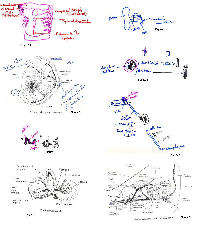
Auricle: margin of 1st visceral cleft.

External auditory canal: Ectoderm of 1st visceral cleft.

Tympanic membrane: -

            Outer epithelial: Ectodermal cleft

            Middle fibrous: Mesoderm.

            Inner Mucosal: Endodermal.

Eustachion tube: Endodermal between 1st and 2nd visceral arch.

Ossicles:

Malleus: Mesoderm of 1st arch 

Incus:  Mesoderm of 1st arch

Stapes:  Mesoderm of 2nd arch.


Inner ear: Ectoderm of hind Brain.


Anatomy: -

External ear:

            Pinna

            External acoustic meatus.


Pinna:


External acoustic meatus:


Tympanic membrane:


Middle ear cleft: consists of ossicles, Eustachion tube, tympanic cavity, aditus ad antrum, mastoid antrum and mastoid air cells.


Tympanic Cavity: biconcave disc


Aditus ad antrum:

lead from epithympanum to mastoid antrum.

Mastoid antum.

Mastoid air cells.


Inner ear: Consists of two parts Osseus part and membraneous part.

The membranous part is surrounded by perilymph and contains endolymph.

The function of the cochlea is related to hearing.

The function of the semicircular canals is related to the control of balance.


Investigation of ear diseases:


History:

  1. Hearing loss (deafness) commonest.

Types:

Conductive

from outside to inside, congenital atresia of the external ear canal, ear wax, tumours of the external canal, perforated ear drum, otitis media with effusin, otosclerosis.

            Sensory neural

                        Congenital hearing loss mainly runs in close family marriage.

                        Trauma to the inner ear.

                        Viral infection like mumps.

                        Noise related hearing problems to those who work in factories for long

times.

Tumours of  the vestibulo cochlear nerve like acoustic neuromas and cerebellar brain tumours.              


  1. Discharge (Otorrhoea)

Serous: like otitis externa

Mucopurulent: like middlke ear disease.

Intermittent: in atticoantral disease.

Continuous: In tubotympanic disease.


  1. Pain:

Local due to otitis externa or otitis media.

Referred could come from nasopharynx, oropharynx, hypopharynx and larynx and it is very common to have ear ache after tonsillits and tonsillectomies.


  1. Itching: Mainly in otitis externa.


  1. Tinniteus:

Subjective sensation of sound in the ear.

Hissing common.

Pulsetile mainly due to vascular abnormalities like A.V fitula in the neck or glomus tumour in the ear.

            Classification:

                        Without any other symptoms., could be stress related.

                        As part of ear disease, mainly in those who have noise induced hearing

loss.

As part of neurological disease due to brain lesions such as Multiple Sclerosis or brain tumours.

                        Accompanied with sytemic diseases:

                                    Renal and cardiac

                        Drugs: Salicylate, ototoxic antibodies.


  1. Vertigo:

Peripheral: due to inner ear diseases like Meniers disease, which is Characterised by fluctuant sensory hearing loss, tinnitus and vertigo. Also infection of the inner ear like bacterial or viral infection could be a cause.

Central: mainly due to brain lesions. It could be vascular or neoplastic.


Examination:

Use good light source

Inspection: pinna, mastoid, side of the face for possible facial palsy.

Otoscopy:

            Pull the pinna upward, outward, backward, with 2x magnification otoscopy.

What to see


Examine the face, XII movement.

Examine the post nasal space (nasopharynx).


Investigation:


Blood tests: FBC, serology tests, auto antibody, also viral titre to check diseases which could cause sensory neural hearing loss.

Hearing test: Audiogram:


Others:

Speech audiometry, electrocochleography, brain stem electric response.

Vestibular function tests:


Radiological:

Plain film.

C.T scan.

MRI scan.


ear.jpg


Ear Diseases


Auricle (pinna):-

Congenital: accessory auricle, microtia, anotia.

Traumatic: Haematoma.

Infectious: Perichondritis

Neoplastic:

Benign

Malignant: BCC, SCC


Ex. Auditory Meatus:-

Congenital: atresia.

Impacted wax, F.B.

Tumours:

                        Benign:

                                    Osteoma

                        Malignant:

                                    Squamous carcinoma.

                                    BCC.

                                    Adeno carcinoma.

Inflammatory:

Otitis externa:

                        Localised: furunculosis caused by staphylococcus aureus.

                        Diffuse:

                                    Bacterial

                                    Fungal:

                                                Aspergellus

                                                Candida

                                    Viral

Symptoms:

                        Pain

                        Discharge


Treatment:       

                        Pain killer.

                        Suction clearanceà to the ear.

                        Antibiotic wick with steroid ointment.

                        In severe cases oral and I.V antibiotics.


Necrotising otitis externa (malignant)

Organism: pseudomonas.

Characterised by spread to the bone causing ostitis cranial nerve palsy, intracarnial spread of infection.

Occurs in autoimmune compromised patients.


Treatment:

                        I.V antibiotics: ciprofloxacullus, painkillers.

Wicks.

                        Debridement


Middle ear disease:

Infections:

                        AOA

                        COM

Acute otitis media:-

Incidence: 16-24 month is the highest.

Actiology: wide horizontally placed Eustachian tube.

1.      More prone to URTI

2.      Enlarged adenoid tissue affects the drainage of the ear.


Bacteriology:

                        Streptococci

                        Staphylococcus, H. influenza.


Clinically:-

Infant:

Pyrexia and screaming.

Older children:

                                    Pain

                                    Deafness.


On examination the drum is dull grey, absent light reflex congested drum and dilated blood vessels.


Investigation:-

                        PTA: conduction loss.


Treatment:-

                        Analgesia

                        Antibiotics: Amoxil orally or I.V.


Chronic otitis media:

            Chronic suppurative otitis media (CSOM)

            Otitis media with effusion (OME)


    CSOM

Complication of AOM with persistent perforation in the drum.

Types:

Turbo-tympanic due to persistent infection through the Eustachian tube causing middle ear problems.


Attico antral disease Due to the development of a retraction pocket in the pars flaccida which keeps enlarging and with keratin inside (Cholesteatoma) The enzyme secreted by this cholesteatoma erodes the bone and causes complications.


Clinically: tubotympanic

Discharge

                        Mucopurulent

                        Persistent.

Deafness: conductive.

O/E: central anterior or posterior perforation.

Nasal examination, post nasal space examination is also examinated.


Investigation

                        Swabs for culture and sensitivity.

Radiology CT scan is helpful in showing bony erosion and the extent of the disease.


Treatment:-

1.      Treat URTI, nose, PNS problems if any.

2.      Suction clearance to the ear under the microscope.

3.      Antibiotics ear drops with steroids.

4.      Surgery: tubotympanic: closed perforation and ossicular repair. (tympanoplasty).


Attico antral disease: mastoid exploration to get rid of the disease.


Otitis media with effusion (OME) (Glue ears)

Incidence: mainly children under the age of 10.

Aetiology: Unknown.

Possible causes:

                        Eustachian tube problems.

                        Viral theory with URTI.

                        Adenoid enlargement.

                        Cleft palate.


Pathology:

Cytology:

Exudate

                        PMNL, macrophage.

Cell debris and biochemically it is viscid fluid contains glycoprotein, nucleoprotein.


Clinically:-

Symptoms:

                        Hearing loss

                        Earache

Signs:

                        T.M straw coloured and dull.

                        Air bubbles, fluid level.

                        Indrawn of T.A


Investigation:

                        PTA: Conductive HL

                        Tympanometry: flat


Treatment:

                        In established cases if it didn't resolve spontaneously.

                        Myringotomies ± grommets and adenoidents.

                        2nd grommet: 35%

3rd grommet: 11%


Complications of otitis media

Rare nowadays due to Antibiotics.

Spread:

                        Superiorly: middle cranial fossa.

                        Posteriorly posterior cranial fossa.

                        Medially: labyrinth.

                        Inferiorly: spread to the jugular bulb.


Extra cranial complications

Mastoiditis

                        Acute

                        Sub acute

                        Chronic

Petrositis.

Labyrinthitis.

Facial nerve paralysis.

Ossicular erosion.


Intra cranial complications:

                        Extra dural abscess.

Sub dural abscess.

Lateral sinus thrombosis

Meningitis

Brain abscess:

                        Temporal.

                        Cerebellar.


Mastoiditis:

Extension of the infection to the mastoid antrum into the mastoid air cells.

Clinically:

Symptoms:

                        Pain behind the ear

                        Deafness

Signs:

                        Tenderness over mastoid antrum.

                        Displacement of pinna downward and outward.

Narrow of the external meatus.

Congested eardrum.


Investigation:

Haematology:

                        Increased white cell count.

                        Radiology:

                                    Plain X-Rays blurring of the cellular mastoid.

                                    CT scan may be indicated for possible complications.


Treatment:

                        I.V antibiotics

                        Pain killer

                        Surgery - mastoidectomy


Otoscelerosis:


Definition: localised disease of optic capsule.

Aetiology:

                        Hereditary tendencies in certain families.

Age: 18-30.

Pathology:

Normal bone replaced by vascular spongy osteoid tissue later it becomes less vascular.

Site: Anterior margin of oval window (commonest).

Clinically:

                        Conductive deafness or mixed deafness

Tinnitus.


O/E:

normal ear drums 90%

active disease (pink-bluish) drum 10%


Treatment:-

                        Stapedectomy (insert prosthesis).


Inner ear disease:


Sensory neural deafness:

Congenital:

Acquired:

Infections

                                    Viral: mumps, measles, herpes zoster oticus.                                                                              Bacterial: after otitis media, syphilis

Presbyacusis

                        Age related deafness


Ototoxic:

                        Aminoglycosides

            Cytaxoic:

                        Salicylate in high dose

Sudden deafness:

                        90% aetiology unknown.


Treatment:

                        Hearing aid.

                        Cochlear implant.


Vertigo:

            Definition: hallucination of movement.

Causes:

Peripheral.

                                    Central.


Peripheral:

Benign paroxysmal positional vertigo:

Attack of the vertigo is related to head movement, due to deposit of on the Cupula of Posterior semicircular canal.

Treatment: positional test Epply manouveure.


Vestibular Neuronitis

                        Proceed by viral URTI with fibrile illness


Treatment: symptomatic.


Meniers disease:


Character:

                                    Deafness: fluctuant SNHL

                                    Vertigo

                                    Tinnitus

                                    Nausea and vomiting


Pathology:

                                    Dilatation of the endolymphatic components (hydropes).

                                    When it rupture potassium is released and causes vertigo.


Aetiology:

                                    Unknown.

                                    Allergy, viral, biochemical theory.


Clinically:

                                    Sudden vertigo.

                                    Fullness of the ear         .


O/E:

                                    Nystagmus (peripheral in type)


Investigation:

                                    PTA : SNHL as the disease progresses.

                                    Vestibular tests: hypo function


Treatment:

                                    Bed rest

                                    Nausea and vomiting: stemetil.

                                    Surgery: if vertigo becomes disabling