CYBERMEDICS
                                                                         © 1999, Venkatesh.K.S

GENE THERAPY
LOGO

PHARMACOKINETICS OF DNA BASED THERAPY

In principle, the pharmacokinetics of DNA-based therapy is no different from conventional drug therapy, i.e., it is an input-output problem involving delivery of DNA to its site of action that subsequently results in up- or down-regulation of a gene product. Accordingly, conventional pharmacokinetic approaches are generally applicable to the delivery system itself (antisense oligonucleotide, DNA-plasmid/vector) and the expressed protein. However, details of the intermediary "black box" are even murkier than usual ("and then a miracle happens") and unlikely to be tractable by classical/pharmacokinetic paradigms. The main reason for this is that the critical events are mostly at the cellular level whereas conventional drug therapy is generally based on whole body pharmacokinetics as reflected by plasma levels. Few studies have addressed the former area, hence, virtually all clinical studies, especially those involving vectors, have been entirely empiric with little or no understanding or consideration of possible pharmacokinetic determinants. As the problems of in vivo studies, in contrast to in vitro investigations, are becoming more appreciated, greater attention is being focussed on such factors.

Antisense Oligonucleotides

Antisense oligonucleotides are 6-60 base polymers with sequence complementary to a selected nucleic acid which hybridize to

(i) mRNA causing sequence specific inhibition of translation and/or RNAse H degradation;
(ii) double-stranded genomic DNA leading to interference in transcription;
(iii) trans-acting elements controlling regulation.

Phosphodiester oligonucleotides are very unstable in blood - t½ is about 5 min. As a result, many backbone modifications have been investigated to increase stability, and most effort has been directed towards nuclease-resistant phosphorothioate (PS) oligonucleotides, i.e., the non-bridging phosphate bound oxygen is replaced by sulfur. "Hybrid" PS-oligonucleotides where a number of 3¢- and 5¢ nucleotides are further modified and "dumbbell", i.e., linked double stranded sequences, have more recently been investigated, because such strategies further stabilize the oligonucleotide.

Oligonucleotides (<5000 kDa) are transported across the capillary bed, with the exception of the brain and gonads, and interact with saturable and specific binding sites on cell surfaces, the number of which depends on tissue type; they are also more abundant on dividing cells. Endocytotic internalization occurs with intracellular distribution to endosomes (non-bioavailable) and also to the nucleus (bioavailable).

After intravenous administration, initial distribution is very rapid (t½ up to 20 min) with plasma elimination half-lives of 15 to 50 hr or more in most animal species based on radioactivity. Distribution of oligonucleotides generally favors the kidney, liver, lung and spleen, and the elimination half-lives from individual tissues may vary. With more stable oligonucleotides, the majority of the measured plasma/tissue level represents the intact agent; however, backbone degradation does occur (kidney and liver). Both unchanged and degraded oligonucleotide are excreted in urine.

While the oligonucleotide field is still in its infancy, the disposition and pharmacokinetic aspects appear to be mainly analogous to those employed with conventional drugs (Agrawal et. al., 1995; Zhang et. al., 1995). Future progress will probably come in the areas of tissue selective delivery, optimizing intranuclear bioavailability, and understanding of the degradation mechanism(s).

DNA-Vector Systems

Currently, there are virtually no data on the in vivo pharmacokinetics of DNA when delivered to cells by viral vectors, liposomes or other delivery strategies. Translational studies from in vitro cultured cells to whole animal studies, including those in humans, have been empirical and mainly designed to test the feasibility of the principal rather than critical evaluation of the problems of optimal formation of a gene product.

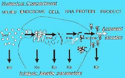
Contemporary strategies for gene therapy focus on either the direct injection of DNA into specific organs or formulating DNA with ligands or carriers that target the DNA to specific cells within the body. When DNA vectors are used like conventional medicines, the DNA is not expected to integrate into the host cell chromosome or to be replicated. Rather, the DNA is intended to reside transiently in the cell as an extrachromosomal (episomal) element, during which time mRNA will be transcribed and a gene product formed. As the DNA is eliminated from the cell, new production of the protein ceases (c.f., strategy of stable and permanent integration into genome by ex vivo approaches). The attraction of using genes as conventional medicines, is that the level of the therapeutic product may be controlled by adjusting the dose and the schedule of administration of the gene. Moreover, the duration of action may be also controlled by the design or formulation of the expression vector. Although these concepts are analogous to those with conventional drugs, the involved pharmacokinetic considerations are novel since they depend on intrinsic intracellular biochemical events such as DNA transcription and mRNA translation rather than the behavior of the "drug" itself. Several factors are likely to be involved.

FIG
IMAGE10
  1. Distribution and extracellular elimination of DNA after in vivo administration.
  2. Efficiency of DNA uptake into cells.
  3. Compartmentalization of DNA within the endosomal, cytoplasmic or nuclear compartments of the cell.
  4. Rate of degradation of DNA within the cell.
  5. Rate of transcription of RNA from DNA.
  6. Stability of the mRNA.
  7. Rate of translation of the mRNA to create the new gene product.
  8. Intracellular compartmentalization or secretion of the gene product.
  9. Pharmacokinetics of the gene product in the body.

Over 98% of the methylmalonyl CoA mutase gene was taken up by the liver using an asialo-glycoprotein/DNA complex with approximately 106 copies/cell. The vector sequence declined with a half-life of 1 to 1-5 hr. Gene expression measured by enzyme activity was greater than control from 6 to 48 hr with a maximum increase of about 30 to 40% (Stankovics et. al., 1995).

With an HLA-B7/cationic liposome vector, intact plasmid DNA by Southern blotting was present in selected tissues at 1 hr (bone marrow, heart, kidney, liver, lung, spleen and muscle representing about 250-16,000 copies/mg genomic DNA, but this was about 20 times less after 24 hr. PCR-detectable plasmid indicated DNA present at 7 and 28 days post-injection. No detectable HLA-B7 protein expression was observed at any time point (Lew et. al., 1995).

     PAGE 1  2  3  4  5  6  7                                                                                                                            Next page


EXTRAS                                MYHOME                                LINKS                                  SEARCH

SEND MAIL SITE MAP