|
(Current Medical Practice (May 1979): 23, 5) ROLE OF LIV.52 IN HEPATITIS AND CIRRHOSIS OF THE LIVERK.K. MALIK , F.R.C.P. (Edin.), D.T.M.H. (Lond.), M. A. M. S. (Ind.),Associate Professor & Head of Gastroenerology Unit and M.B. PAL, M. B. B. S. (Cal.), Research Scholar, Gastroenterology Unit, Department of Medicine, Institute of Post-graduate Medical Education and Research, Calcutta - 700 020. The liver plays a key role in the metabolic activity in the human body. Although its structure is simple, its functions are most complex. Even to the present day, diseases of the liver have continued to remain an enigma. Liver is actively concerned with many vital productive, synthesizing and regulatory functions. It participates in many metabolic functions, including detoxification. During the process of detoxification the toxin may bring about the death of some of the cells of a liver lobule or lobules or cause mild but repeated damage, continuously or intermittently. It is difficult to exactly evaluate and assess disturbance of liver function and correlate it with the degree of damage since the liver has great reserve power and at any given time it does not work at full capacity. However, today, clinical evaluation and its correlation with the biochemical and histopathological changes in the liver combined with hepatic scinti-scanning offers a comprehensive approach to the study of the problem. Different workers have reported about the efficacy of Liv.52 in cirrhosis of liver (Mukherji and Dasgupta, 1971 and Mehrotra and Tandon, 1973 and in hepatitis (Sharma et al 1974). In animal experiments, Liv.52 is found to increase the albumin production in the liver (Sheth et al 1960) while Mukerji and Dasgupta, 1970 pointed out its efficacy as a hepatic stimulant. But the probable mechanism leading to improvement of the liver function still remains to be elucidated. Liv.52 is a compound of indigenous drugs reputed to have their effects on hepatic function. It is claimed to have protective and regenerative effect on hepatic parenchyma and have anabolic and diuretic action and no acute or chronic toxicity. Each tablet of Liv.52 (The Himalaya Drug Co.) contains:
and is prepared in the juices and decoctions of various hepatic stimulants. In this study, 20 cases of Infective (Viral) Hepatitis have been taken as representative of acute hepatic damage and 122 cases of Cirrhosis of liver as of chronic hepatic damage. LIV.52 IN ACUTE HEPATIC DAMAGE This group consisted of 20 cases of viral hepatitis. These were divided into two groups. One group of 10 selected alternatively from hospital admissions for viral hepatitis was given Liv.52 tablets 2 q.i.d. and was designated as Liv.52 group. The other group of 10 served as a control group. Both groups were given the same routine treatment and resuscitative measures as were necessary. In the test group, Liv.52 tablets 2 q.i.d. were administered for two months. Clinical and biochemical assessment of each case was noted initially and at the end of 2, 4 and 8 weeks of the study. The symptomatology and its progress and also the laboratory and the biochemical progress in the Liv.52 group and the control group are presented in Tables I and II.
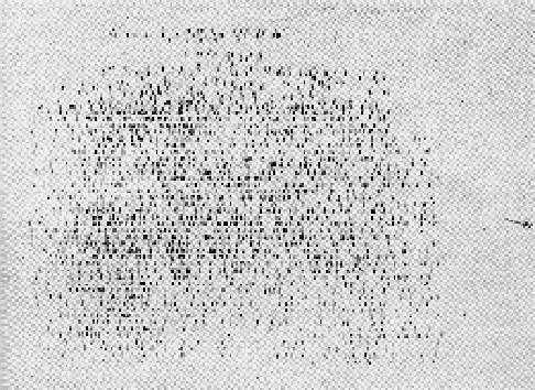
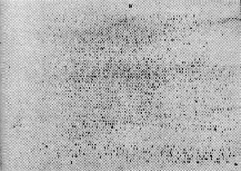
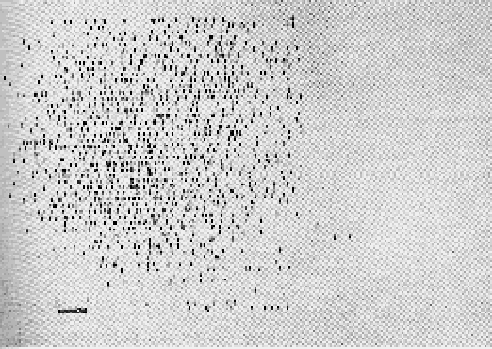
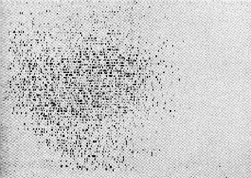
From Table I it is evident that jaundice, anorexia, hepatomegaly and fever improved earlier in Liv.52 group, thereby showing a definite clinical response. LIV.52 IN CHRONIC HEPATIC DAMAGE : STUDIES WITH SCINT-SCAN Eight cases were studied in this group. During the course of the study, a detailed history of each case was taken and the presenting symptoms and signs were noted initially and at the end of 4 and 8 weeks and the trial was concluded at the end of 12 weeks. Clinical and biochemical assess-ment of all the cases was done before and after the therapy. Each patient received 2 tablets of Liv.52 q.i.d. for 3 months. Radioactive Rose Bengal I-131 liver scan was done before and after the Liv.52 study lasting 3 months. All eight cases of cirrhosis were given 120 p-curie of radioactive Rose Bengal I-131 dye after which scinti-scan of liver with Magnus Scanner Machine was done. A coloured hepatic excretion scan was obtained in the anteroposterior (AP) and right lateral (RL) views. Excluding the blue colour as "background activity" the liver size was determined each time by tracing out the area of the green, yellow and red colour on squared paper outlining the mass of functioning liver tissue in square centimetre as projected in the liver scan. This seems to be one of the most promising simple and painless (though costly) methods of evaluating the efficacy of drug action on the total mass of functioning hepatocytes by comparing the magnitude of Rose Bengal excretion in the pre and post-treatment scans.
The control group was made up of 34 cases selected alternately from the hospital admissions for cirrhosis. This group received the usual treatment for cirrhosis including diuretics and blood transfusion which where necessary but not Liv.52. The other 34 alternate cases along with further 46 consecutive admissions for cirrhosis formed the Liv.52 group. The 80 cases in this group received Liv.52, 2 tablets q.i.d. for 3 months in addition to the routine treatment for cirrhosis. Detailed history and clinical examination were done in each case, clinical and biochemical assessment was done on admission at 4, 8 and finally at 12 weeks of the therapy. Special studies included hepatic function tests by universally accepted techniques. These included total blood billirubin, blood proteins, enzyme studies S.G.O.T., S.G.P.T., serum alkaline phosphatase and 45 minutes bromsulphthalein dye clearance. Oesophagogram was done in 37 cases and splenoportal venogram in 20 cases in the control group and in Liv.52 group, both oesophagogram and splenoportal venogram were done in 80 and 40 cases respectively. Hepatic histopathology using vim-Silvermann needle was done to establish the diagnosis of cirrhosis in each case but this being both a blind and painful process, its repetition was not possible except in 6 cases.
In the control group, out of 34 cases, 30 showed evidence of cirrhosis with pseudo-lobule formation. Splenoportal venography was possible in 20 cases and it showed evidence of intrahepatic obstruction and or collateral branches in 16 cases and a normal venogram in 4 cases. Oesophagogram showed positive evidence of oesophageal varices in 22 cases. In the Liv.52 group, of 80 cases of cirrhosis liver, 74 cases were histologically positive for cirrhosis. Splenoportal venography was done on 40 cases and 30 (75%) showed evidence of either intrahepatic obstruction and/or collateral’s from the splenic vein. Oesophagography was done in all 80 cases and out of these 53 (66%) had evidence of oesophageal varices. On repeat liver biopsy in 6 cases, 3 showed some improvement compared to the first biopsy findings but as this is blind process, no definite conclusions can be drawn from this. Of the either cases of cirrhosis on whom scinti-scan of the liver was done, scan pictures of case Nos. 1, 3, and 5 are reproduced on pages 5, 6 and 7.
These pictures show improved function of hepatic cell mass after treatment with Liv.52. CONCLUSION In our study of viral hepatitis cases, jaundice improved in 60% of the cases in the control group whereas it improved in 80% of the cases in the Liv.52 group within 2 weeks. Jaundice had cleared in both groups at the end of 4 weeks. Besides jaundice, the symptoms of anorexia, fever and hepatomegaly were relieved earlier in the Liv.52 group than in the control group. After two weeks, there was a decrease in serum albumin in 10% of the cases in the Liv.52 group whereas in the control group decreased albumin was found in 20% i.e. after two weeks’ Liv.52 therapy there is less decrease in serum albumin. There was hardly any change in serum globulin and alkaline phosphatase levels following treatment. However, there was marked improvement in S.G.O.T., S.G.P.T. at 2nd and 4th week with Liv.52. Transaminase activity was normal in 80% of the cases in two weeks and in all the cases within 4 weeks on Liv.52, whereas without treatment only 50% and 80% of control cases attained normalcy in the same period. In conclusion, we wish to point out that Liv.52 therapy is usually effective both symptomatically and biochemically in two weeks. The maximum effect of the treatment was evident in anorexia, jaundice, hepato-megaly, serum bilirubin and transaminase levels. Symptomatic relief was seen in 90% of the cases whereas biochemical values returned to normal in about 30% of the cases. Out of the 8 cases of cirrhosis who underwent the liver scan both before and after treatment, 5 cases (63%) showed improvement in the area of functioning hepatocytes. 3 cases (37%) did not show any improvement in the scan area even after treatment. But the antero-posterior and the right lateral views of one of these 3 cases and the area of antero-posterior view in another case showed hardly any change from pre-treatment to post-treatment values. From the above observations, it is clear that 63% of the cirrhotic cases showed improvement in the total mass of functioning hepatocytes following treatment with Liv.52 whereas in another 25% of cases there was hardly any perceptible difference in one of the views in the pre- and post-treatment scan areas. There was marked symptomatic and biochemical improvement after treatment with Liv.52. Symptoms of haematemesis and clinical jaundice and signs of hepatic failure disappeared in all cases within 4 weeks’ therapy with Liv.52. The Radioactive Rose Bengal 131 test reflects better hepatocellular function than other relevant liver function tests. The third phase of our study consisting of 114 cases of cirrhosis showed comparable signs and symptoms in both the Liv.52 and control cases, but treatment with Liv.52 showed definitely better improvement in ascites, oedematous feet and prominent abdominal veins. This indicates that Liv.52 therapy has a distinctly beneficial effect on fluid accumulation in cirrhotic cases. Regarding other symptomatology the improvement was only marginal after treatment. Biochemically, when compared to control cases, there was significant improvement in S.G.O.T. level and B.S.P. clearance rate in the Liv.52 group. The beneficial effect on S.G.O.T. level may be ascribed to increased mass of functioning hepatocytes. REFERENCES
|
||||||||||||||||||||||||||||||||||||||||||||||||||||||||||||||||||||||||||||||||||||||||||||||||||||||||||||||||||||||||||||||||||||||||||||||||||||||||||||||||||||||||||||||||||||||||||||||||||||||||||||||||||||||||||||||||||||||||||||||||||||||||||||||||||||||||||||||||||||||||||||||||||||||||||||||||||||||||||||||||||||||||||||||||||||||||||||||||||||||||||||||||||||||||||||||||||||||||||||||||||||||||||||||||||||||||||||||||||||||||||||||||||||||||||||||||||||||||||||||||||||||||||||||||||||||||||||||||||||||||||||||||||||||||||||||||||||||||||||||||||||||||||||||||||||||||||||||||||||||||||||||||||||||||||||||||||||||||||||||||||||||||||||||||||||||||||||||||||||||||||||
