3.1
Normal MRI anatomy of the Brain
The
human adult brain is one of the largest organs of the body and can be
divided into four main principal parts: brain stem, diencephalon,
cerebrum and cerebellum. The
brain stem consists of the medulla oblongata, pons and midbrain and is
continuous with the spinal cord. Above
the brainstem is the diencephalon, consisting primarily of the thalamus
and hypothalamus.
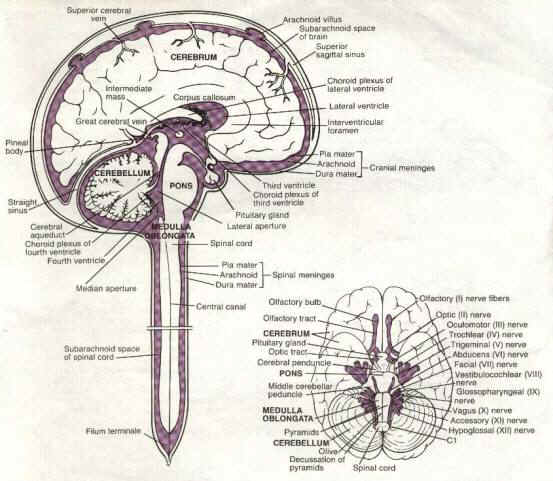
Diagram
1 Sagittal section of the brain and spinal cord (adapted from Snell,
1995).

The
cerebrum occupies most of the cranium and spreads over the diencephalon.
The cerebrum is made of two halves the right and left cerebral
hemispheres, which are further, divided into lobes: frontal, parietal,
temporal and occipital. The
cover of each hemisphere is called the cortex and is composed of gray
matter, while the underlying myelinated axons are composed of white
matter. Inferior to the to
the cerebrum and posterior to the to the brain stem is the cerebellum
(Snell, 1995; Tortora and Grabowski, 1993).
Grey and white matter contrast, and contrast within the different
white matter regions is far greater on MRI than any other imaging
modality. A major
contribution to this contrast is myelin density, which is by far greater
in white matter. Also, the
water content of gray matter is greater than that in white matter.
Due
to the lower water content of white matter, it will have a very short T1
and T2 relaxation times than that of gray matter.
On T1-weighted images gray matter will have an intermediate
density (gray) and darker than white matter.
On T2-weighted images the signal intensities tend to be reversed
i.e. the white matter appears darker than the gray matter (Woodward,
2001).
The
cranial bones and the cranial meninges protect the brain where the outer
layer is the dura mater, the middle arachnoid and the inner is the pia
mater. Bone, such as the
cranial bones, will appear dark on T1 and T2-weighted images of the
brain. However, beam-hardening artefacts of CT when imaging through
the petrous region are not present in MRI.
Calcification within the brain, as a calcified choroid plexus or
basal ganglia will also appear dark on T1 and T2-weighted images
(Woodward, 2001).
The
two cerebral hemispheres are separated by an extension of the dura mater
referred to as the falx cerebri. The
falx cerebelli and the tentorium cerebelli are also an extension of the
dura mater where the former separates the cerebellar hemisphere and the
latter separates the cerebrum from the cerebellum.
The brain is nourished and protected against chemical or physical
injury by cerebrospinal fluid (CSF), which circulates through the
subarachnoid space and the four ventricles within the brain.
Figure
1 Mid-Sagittal T1-Weighted MR image

1=rostrum of corpus Callosum, 2=genu of Corpus Callosum, 3=body, 4=splenium, 5=cingulategyrus, 6=gyrus rectus, 7=precentral gyrus, 8=postcentral gyrus, 9=frontal lobe, 10=occipital lobe, 11=lateral ventricle, 12=Fornix, 13=thalamus, 14=third ventricle, 15=mammillary body, 16=cerebellar peduncle, 17=pons, 18=medulla, 19=vermis of cerebellum, 20=fourth ventricle, 21=parietal lobe, 22=tentorium cerebelli
Figure
2 Proton Density MR image showing the basal ganglia.

1=gray matter, 2=white matter, C=caudate nucleus, P=putamen, G=globus pallidus, I=internal capsule, T=thalamus.
Diagram 2 The ventricles (adapted from Snell, 1995)

The
marked difference in relaxation times between brain and water enables the
demonstration of these structures surrounded by CSF. CSF has a high proton density and appears with a very low
signal on T1-weighted images and bright on T2-weighted images (i.e. TR of
2000+) (Westbrook, 1999). Nevertheless,
the appearance of CSF in FLAIR sequences is dark even though it is a
heavily T2 weighted image since the signal from CSF is voided without
affecting interparenchymal tissues (Woodward, 2001).
Fat is considered as another important benchmark in MRI. Since it has a very short T1 relaxation time it will appear bright on T1-weighted images. It has intermediate signal intensity on T2-weighted images and appears gray. The appearance of fat on T1-weighted images can sometimes be problematic in post contrast imaging if the precontrast is not formed first. This is especially critical in the pituitary fossa, orbits and the base of skull, where both fat-containing areas and enhancing areas will appear identical on T1-weighted images. This problem has been solved with the introduction of fat suppression enabling identification of the enhancing lesion (Woodward, 2001).
Figure 3 T1-Weighted coronal section at the level of 3rd ventricle


The brain is also well supplied with nutrients and oxygen mainly by the anastosmosis between the right and left internal carotid arteries, their branches and the posterior cerebral arteries, which form the Circle of Willis. However, this fast flowing blood will appear dark or black on T1 and T2-weighted spin echo images of the brain. The blood supply to the brain can be well demonstrated on MRI by Magnetic Resonance Angiography (MRA) either by using time of flight (TOF), phase contrast MRI or Gadolinium enhanced MRA (Jager et al, 2001; Ryan and McNicholas, 1994; Tortora and Grabowski, 1993). MRA is discussed in more detail in section 9.