CASE PRESENTATION AND DISCUSSION ON ABDOMINAL MASS
OLIVER S.LEYSON
Surgery Resident
Department Of Surgery
Ospital Ng Maynila Medical Center
General Data
6 mos, female from Sta Ana Manila
Chief Complaint
Abdominal Mass
History of Present Illness
4 mos PTA -------------------------à mother noted abdominal enlargement with
abdominal mass, no change in bowel habits
(-) vomiting. No consult done
No medications taken
no other symptom noted
2 weeks PTA ----------------------à progression of abdominal
enlargement, poor suck,
Prompted consult at private MD
advised consult OMMC
1 day PTA ----------------------- à persistence of abdominal enlargement,
associated with vomiting refusal to feed prompted consult OMMC
Admitted
Past Medical History
No previous hospitalization
Family History
Unremarkable
Personal Social History
Gestation: patient was born to a 26 yo G1P1
(+) prenatal check up at health center
(+) intake of Multivitamins and Ferrous sulfate
(-) maternal illness (-) exposure to radiation
(-) intake of teratogenic drugs
Delivery: delivered term via NSD at OMMC
(-) fetomaternal complication
Neonatal- good suck, good cry, no retractions, no jaundice.
Immunization- (+) BCG, (+) DPT2 (+) OPV2
Feeding- Breastfed since birth up to present
Growth and development- at par of age
Physical Examination
General: Awake, comfortable, poorly-nourished, fairly developed, not in cardio-
respiratory distress
CR: 126/min RR: 40/min T: 36.7C BW: 6kgs
HC: 42 cms CC: 40 cm AC: 48 cm BL: 64 cm
HEENT: normocephalic, pink palpebral conjunctiva anicteric sclerae, (-) NAD
Chest and Lungs: Symmetrical chest expansion, no retraction,clear breath sounds.
Heart: Good heart tone, normal rate regular rhythm no murmrur.
Abdomen: Globular, tensed shiny, bulging flank mass extendingto the anterior the
abdominal wall right measuring 10 x 10 cm solid, non hard, firm, non-
movable, non tender,
Extremities: grossly normal full equal pulses
Rectal Exam: no skin tags, no fissures, good sphincteric tone, no mass, no tenderness,
no bleeding, with yellowish stool on examining finger.
Salient Features
- 6 mos old/ Female
- gradually enlarging abdominal mass ( 6 mos duration)
- abdominal mass 10x10cm, firm, non hard, non-movable extending to the right anterior abdominal wall
- No change in bowel habits
- Asymptomatic
- Female
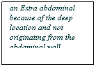
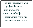
Algorithm for abdominal mass
Abdominal Mass
Extra-abdominal Intra-abdominal
Retroperitoneal Intraperitoneal

Clinical Diagnosis
|
|
Diagnosis |
Degree of Certainty |
|
Primary Clinical Diagnosis |
Abdominal Mass probably retroperitoneal |
60 % |
|
Secondary Clinical Diagnosis |
Abdominal Mass probably intraperitoneal |
40% |
Paraclinical diagnostic procedure:
• Do I need a paraclinical diagnostic procedure?
• Yes, because I’m not yet certain of my primary clinical diagnosis.
Goal
• Identify the organ of origin and the extent of the tumor
PARACLINICAL DIAGNOSTIC PROCEDURE
|
|
BENIFIT |
RISK |
COST |
AVAILABILITY |
|
X-RAY |
+ |
Radiation exposure |
150 Php |
Available |
|
UTZ |
++ |
minimal |
500 Php |
Available
|
|
CT SCAN |
++++ |
Radiation exposure |
5000 Php |
Not available |
|
MRI |
++++ |
minimal |
10000 Php |
Not available |
I have chosen CT scan of the abdomen as the procedure that would increase my degree of certainty of my primary clinical diagnosis, because it can demonstrate the organ involved and the extent of the tumor comparable and cheeper than MRI and more sensitivity than that with the x-ray. Ct scan is the most cost effective procedure for this patient
CT Scan result:
![]() Huge homogenous soft tissue mass
Huge homogenous soft tissue mass
![]() Occupying the Right hemiabdomen and extends to the left
Occupying the Right hemiabdomen and extends to the left
![]() displacing the bowels contralaterally and inferiorly.
displacing the bowels contralaterally and inferiorly.
![]() The liver is displaced superiorly and anteriorly
The liver is displaced superiorly and anteriorly
![]() Cystic component located inferior portion and measures about 10 x9
cm
Cystic component located inferior portion and measures about 10 x9
cm
![]() Both kidneys are functioning, displaced inferiorly more at the
right, no hydronehprosis. Spleen unremarkable, filled bowels compressed
towards the left
Both kidneys are functioning, displaced inferiorly more at the
right, no hydronehprosis. Spleen unremarkable, filled bowels compressed
towards the left
Impression: Mass is retroperitoneal in location extra-renal, extra-hepatic


Retroperitoneal Mass
Wilms Tumor Neuroblastoma Teratoma
We can readily rule out Wilms Tumor here in the CT scan result because of the extra renal in origin of the tumor. Neuroblastoma cannot be totally ruled out because it’s the most common solid tumor in children, we can therefore say that with 90% degree of certainty the patient has teratoma.
Clinical Diagnosis
|
|
Diagnosis |
Degree of Certainty |
|
Primary Clinical Diagnosis |
Retroperitoneal Mass probably 2ndry to Teratoma |
90 % |
|
Secondary Clinical Diagnosis |
Retroperitoneal Mass Probably 2ndry to Neuroblastoma |
10% |
PARACLINICAL DIAGNOSTIC PROCEDURE:
• The CT Scan increased my degree of certainty to 90 % basing my decision both on pattern recognition and prevalence so I do not need further a paraclinical diagnostic procedure.
Pre Treatment Diagnosis
|
|
Diagnosis |
Degree of Certainty |
Treatment |
|
Primary Clinical Diagnosis |
Retroperitoneal Mass probably 2ndry to TERATOMA |
90 % |
Surgical |
· Since the treatment to both of my primary and secondary clinical diagnosis are relatively the same, I will proceed now to Treatment Option.
Preoperative treatment goals
Complete extirpation of mass
Achieve locoregional and systemic control of the tumor
Treatment Options
|
Procedure |
Benefit |
Risk |
Cost |
Availability |
|
Surgery |
Complete extirpation and tumor burden ++++ |
Risk of Surgery & anesthesia |
5000 php |
available |
|
Surgery + Chemotheraphy |
Complete extirpation and tumor burden ++++ |
Risk of Surgery & anesthesia Risk of toxicity chemotheraphy |
20,000 php |
available |
|
Chemotheraphy alone |
to down grade the tumor, ++ |
Risk of toxicity chemotheraphy |
15,000 php |
available |
The most cost effective treatment surgical with chemotheraphy option because it achieves our goal of extirpation of the mass and locoregional and systemic control of the tumor.
Pre-op preparation
• Informed consent
• Psychosocial support
• Optimize patient’s health
1. Maintain orogastric tube to decompress the stomach
2. Adequate fluid resuscitation to correct hypovolemia & electrolyte abnormalities
3. Preoperative antibiotics
• Screen for any condition that will interfere with treatment
Operative technique
![]() Patient lying supine under GA
Patient lying supine under GA
![]() Asepsis / antisepsis done
Asepsis / antisepsis done
![]() Sterile drapes placed
Sterile drapes placed
![]() A transverse incision done up to the peritoneum
A transverse incision done up to the peritoneum
![]() Intra-operative findings noted
Intra-operative findings noted
![]() Incision extended up to left upper quadrant
Incision extended up to left upper quadrant
![]() Sharp & blunt dissection of the mass from the subhepatic area
Sharp & blunt dissection of the mass from the subhepatic area
![]() Transverse colon and duodenum dissected from the mass.
Transverse colon and duodenum dissected from the mass.
![]() Dissection carried up to the retroperitoneal area with
electrocautery.
Dissection carried up to the retroperitoneal area with
electrocautery.
![]() Feeding vessels clamped and ligated
Feeding vessels clamped and ligated
![]() Complete excision of mass
Complete excision of mass
![]() complete gauze and instrument count
complete gauze and instrument count
![]() colon reperitonealized
colon reperitonealized
![]() abdominal closure
abdominal closure
![]() - Posterior rectus sheath with Vicryl 2.0
- Posterior rectus sheath with Vicryl 2.0
![]() - Anterior rectus sheath with Vicryl 2.0
- Anterior rectus sheath with Vicryl 2.0
![]() - Skin with Vicryl 5.0
- Skin with Vicryl 5.0
![]() Dry sterile dressing
Dry sterile dressing
Intraoperative findings
![]() 20 x 15 x 15 cm cystic mass with solid components noted at
subhepatic area extending to the splenic flexure
20 x 15 x 15 cm cystic mass with solid components noted at
subhepatic area extending to the splenic flexure
![]() Mass was noted adherent to the transverse colon and to the right
of the duodenum.
Mass was noted adherent to the transverse colon and to the right
of the duodenum.
![]() It extends to the medial side of the right kidney.
It extends to the medial side of the right kidney.
![]() Mass weighed 2 kgs.
Mass weighed 2 kgs.
![]() On cut section we noted 500 cc of straw colored fluid which is
loculated.
On cut section we noted 500 cc of straw colored fluid which is
loculated.
![]() Solid areas heterogenous with some fleshy and some fatty .area
Solid areas heterogenous with some fleshy and some fatty .area


Operation performed
Laparotomy, excision
Final Diagnosis
Retroperitoneal Teratoma
Histopathologic Diagnosis
Immature Teratoma Grade II
Post op Management:
• Maintained on NPO
• Adequate analgesia given
• Antibiotics continued
• Adequate pulmonary support
• Monitoring of early complications
• Start early Feeding as soon as the patient started to soil
• Daily wound care given
• Antibiotics given
Follow up plan
• Continue medications at home
• Continue daily wound care
• Follow up after a week for start of chemotheraphy ( 6 cycles)
– Bleomycin
– Carboplatin
– Etoposide
Aftermath of the patient
• Achieved the following:
– Resolution abdominal mass
– Improved patients condition
– Happy and contented with the outcome
– No medicolegal suit