Photos From Zachary's AMPLATZER® Duct Occluder Placement During
Catheterization >
 What Happens During A Catheterization>
Once your child is taken into the cath lab, your child will be asked to lie down on a large x-ray table. Your child's temperature, heart rate, blood pressure, respirations, and oxygen saturation (SAT) will be monitored continuously during the procedure. A soap called Betadine will be used to wash the areas of the body where the catheters will be inserted. An IV line will be started in a vein in order to give fluids and medications to your child during the procedure. The medications given before and during the cath will cause the patient to sleep during the entire procedure. After your child is asleep, the cardiologist will inject some numbing medication into the areas of the skin where the catheters will be inserted - usually the groin area (the top of the legs), but sometimes the neck, too. After the area is numb, small plastic tubes (sheaths) about the size of a piece of spaghetti are inserted into the large blood vessels. Through these sheaths, long plastic tubes (catheters) are guided into the heart. X-ray helps the doctor guide the catheters into the correct place. Once the catheters are in place, small amounts of blood are taken through the catheters from the various chambers and blood vessels in the heart to determine the amount of oxygen in the blood. The blood pressure in the heart is also measured at the same time. Next, a special dye (contrast) is injected through the catheters, into the chambers and blood vessels of the heart. The dye shows up as a bright white shadow on x-ray film, and allows your doctor to see the shape of the structures and the flow of the blood through the heart and lungs. Motion x-ray pictures are recorded as the dye passes through the heart. If a device is to be used to occlude an unnecessary arterial, venous, or surgically created vascular communication, a catheter is advanced under fluoroscopy from one of the arteries to the communication site. Using the catheter, the device is placed in the vascular communication and blood flow through the communication is blocked. Pictures are taken after the device has been placed to make sure that all blood flow in the communication has been blocked. At the end of the catheterization, the catheters and sheaths are removed, and firm pressure is held to the site for about 10 - 15 minutes. After the bleeding has stopped at the site, a large bandage called a pressure dressing is placed over the puncture site. The pressure dressing helps reduce the chance of bleeding, swelling, and bruising at teh catheterization site. During the time that the cath lab nurses are applying this dressing, the cardiologist will usually find you in the waiting room to inform you of the findings of the catheterization. After the cardiac cath, your child will go to the Recovery Room. Usually one parent is allowed to stay with the child. Once your child is awake enough, he/she will be allowed to take clear liquids. After a couple of hours, your child will be transferred to the Extended Care Unit before being released or be admitted to the Hospital if an overnight watch is needed.
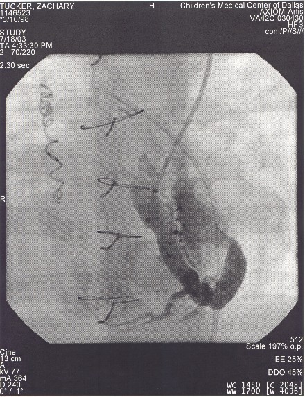 Before The Device Placement:
You Can See A Curly Coil From A Previous Cath, And Four Chest Wires From A Previous Open Heart Closure
You Can Also See The Large Curled Vein That Was Determined To Be Not Needed And Shunting Blood
Before The Device Placement:
You Can See A Curly Coil From A Previous Cath, And Four Chest Wires From A Previous Open Heart Closure
You Can Also See The Large Curled Vein That Was Determined To Be Not Needed And Shunting Blood
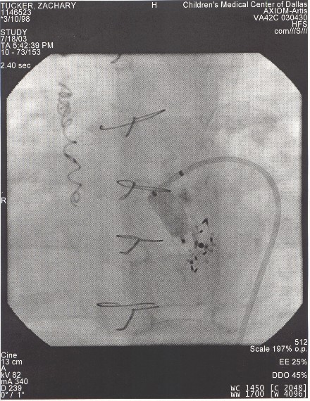 After The Device Placement: The Vein Disappears
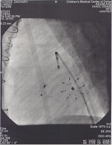 After The Device Placement:
If You Look Closely You Can See The Top Hat Shape Of The Amplatzer Device On The Left And The "Star" Shape Of The CARDIOSEAL Device On The Right
After The Device Placement:
If You Look Closely You Can See The Top Hat Shape Of The Amplatzer Device On The Left And The "Star" Shape Of The CARDIOSEAL Device On The Right