What is Hydrocephalus?
Hydrocephalus is commonly known as 'water on the brain', although this is not accurate. A watery fluid, known as cerebro-spinal fluid (or CSF, for short), is produced constantly inside each of the four spaces or ventricles inside the brain. The CSF normally flows through narrow pathways from one ventricle to the next, then out over the outside of the brain and down the spinal cord. The CSF is absorbed into the bloodstream and the amount and pressure are normally kept within a fairly narrow range. If the drainage pathways are obstructed at any point, the fluid accumulates in the ventricles inside the brain, causing them to swell - resulting in compression of surrounding tissue. In babies and infants, the head will enlarge. In older children and adults, the head size cannot increase as the bones which form the skull are completely joined together.
What causes Hydrocephalus?
The condition is caused by the inability of CSF to drain into the bloodstream. There are many reasons why this can happen:
Congenital Hydrocephalus
This means that hydrocephalus is present at birth. It is important to remember that this term does not imply that it is hereditary. Often the exact cause of congenital hydrocephalus cannot be determined.
Prematurity
Babies born prematurely are at risk of developing hydrocephalus. A baby born early is far more vulnerable than one which goes the full term since many parts of the body will not have matured; for example, the brain is still in a very active stage of development. The area which lies just beneath the lining of the ventricles in the brain is particularly important - because of the activity in this area it has a plentiful blood supply. Its blood vessels are very fragile and can be easily burst if the baby suffers too large a swing in blood pressure or in the amount of fluid in the system.
If these complications do occur, then the baby is at risk of developing a haemorrhage from rupture of the fragile vessels. This can lead to a blood clot developing, which in some cases is big enough to break through the wall of the ventricle. Should the clot block the flow of CSF, the baby will develop hydrocephalus. The blockage may be temporary or permanent.
Spina Bifida
The majority of babies born with spina bifida have hydrocephalus. In addition to the lesion in the spinal cord, there are abnormalities in the physical structure of certain parts of the brain which develop before birth. This prevents proper drainage of the CSF. The increase in pressure due to this can also compress the abnormal parts of the brain even further.
Other forms
of brain haemorrhage, including those occurring in adults, can result in this type of post-haemorrhage hydrocephalus.
Meningitis
This is an infection of the membranes covering the brain. The inflammation and debris from this infection might block the drainage pathways resulting in hydrocephalus. Meningitis can occur at any age.
Tumours
Tumours can be benign or malignant. Tumours of the brain cause compression and swelling of surrounding tissues, resulting in poor drainage of CSF. In the treatment of brain tumours, it is often necessary to include measures to control hydrocephalus, which might only be temporary.
Genetic
In very rare circumstances, hydrocephalus is due to a genetic familial cause - in other words, it is hereditary.
Other causes
There are many other very rare causes of hydrocephalus. There is a particular group of disorders involving the formation of fluid filled cysts (for example, Dandy-Walker cyst) in the CSF system. In these cases, hydrocephalus is often due to pressure on the surrounding tissues by the enlarging cyst.
How is Hydrocephalus Treated?
Some forms of hydrocephalus require no specific treatment. Other forms are temporary and do not require long-term treatment. However, most forms do require to be treated, and this is usually done surgically. Drugs have been used for many years but they may have unpleasant side effects and are not always successful.
The usual treatment is to insert a shunting device. It is important to note that this does not 'cure' the hydrocephalus and damage to the brain tissue remains. Shunting controls the pressure by draining excess CSF, so preventing the condition becoming worse. Symptoms caused by raised pressure usually improve but other problems of brain damage can remain.
Increasingly an operation called Third Ventriculostomy is being performed in specialist units.
What is a Shunt?
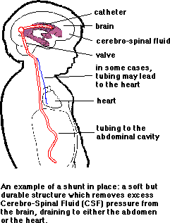
A shunt is simply a drain which diverts the accumulated CSF from the obstructed pathways and returns it to the bloodstream. The device consists of a system of tubes with a valve to control the rate of drainage and prevent back-flow. It is inserted surgically so that the upper end is in a ventricle of the brain and the lower end leads either into the heart (ventriculo-atrial) or into the abdomen (ventriculo-peritoneal). The device is completely enclosed so that all of it is inside the body. The fluid which is drained into the abdomen passes from there into the bloodstream. Other drainage sites such as the outer lining of the lungs (ventriculo-pleural shunt) can also be used. In most cases, the shunts are intended to stay in place for life, though alterations or revisions might become necessary from time to time.
Are there any complications?
Complications are usually caused either by blockage of the system or infection. They are only occasionally due to mechanical failure of the valve. The tube or catheter may become too short as the individual grows and an operation to lengthen it might be necessary.
Symptoms vary enormously between individuals and it is unwise to rely on a list which might not apply in any particular instance. Previous personal experience of a shunt problem is usually a reliable guide as to what to look for.
Shunt Blockage
Symptoms usually develop gradually. In some cases, it shows itself in a gradual deterioration in overall performance. Occasionally, symptoms are quite suddenly severe and may include headaches and vomiting. Various tests can be carried out to confirm the diagnosis. Medical advice should be sought urgently if a shunt blockage is suspected.
Shunt Infection
Symptoms vary with the route of drainage. In ventriculo-peritoneal shunts, the symptoms will often resemble those of a blockage. This is because the shunt becomes infected and the lower catheter is very often sealed off by tissue. There may be accompanying fever and abdominal pain or discomfort. In infection of ventriculo-atrial shunts, fever is present in most cases though often intermittently. Anaemia is frequently present, sometimes skin rashes along with joint pains.
In contrast to ventriculo-peritoneal shunts, such infections sometimes do not become apparent for months after the operation at which they were contracted.
Various tests can be carried out for shunt infection and medical advice should always be sought if an infection is suspected.
How are shunt problems treated?
Shunt blockages which are causing illness usually require an operation to replace or adjust the offending part of the shunt. Shunt infections are usually treated by removal of the whole shunt and a course of antibiotics before insertion of a new system. Modern approaches to antibiotic therapy mean that such treatment can be expected to succeed in most cases.
Links
BACK TO PAGE 1
PAGE 7
PAGE 6:
First page pictures:
Second Page Pictures:
Third Page Pictures:
visitor # 
THE HEART
Atrial Septal Defect
Ventricular Septal Defect